Deposition Date
2011-12-14
Release Date
2012-02-29
Last Version Date
2024-10-16
Entry Detail
PDB ID:
3V44
Keywords:
Title:
Crystal structure of the N-terminal fragment of zebrafish TLR5
Biological Source:
Source Organism(s):
Danio rerio (Taxon ID: 7955)
Eptatretus burgeri (Taxon ID: 7764)
Eptatretus burgeri (Taxon ID: 7764)
Expression System(s):
Method Details:
Experimental Method:
Resolution:
2.83 Å
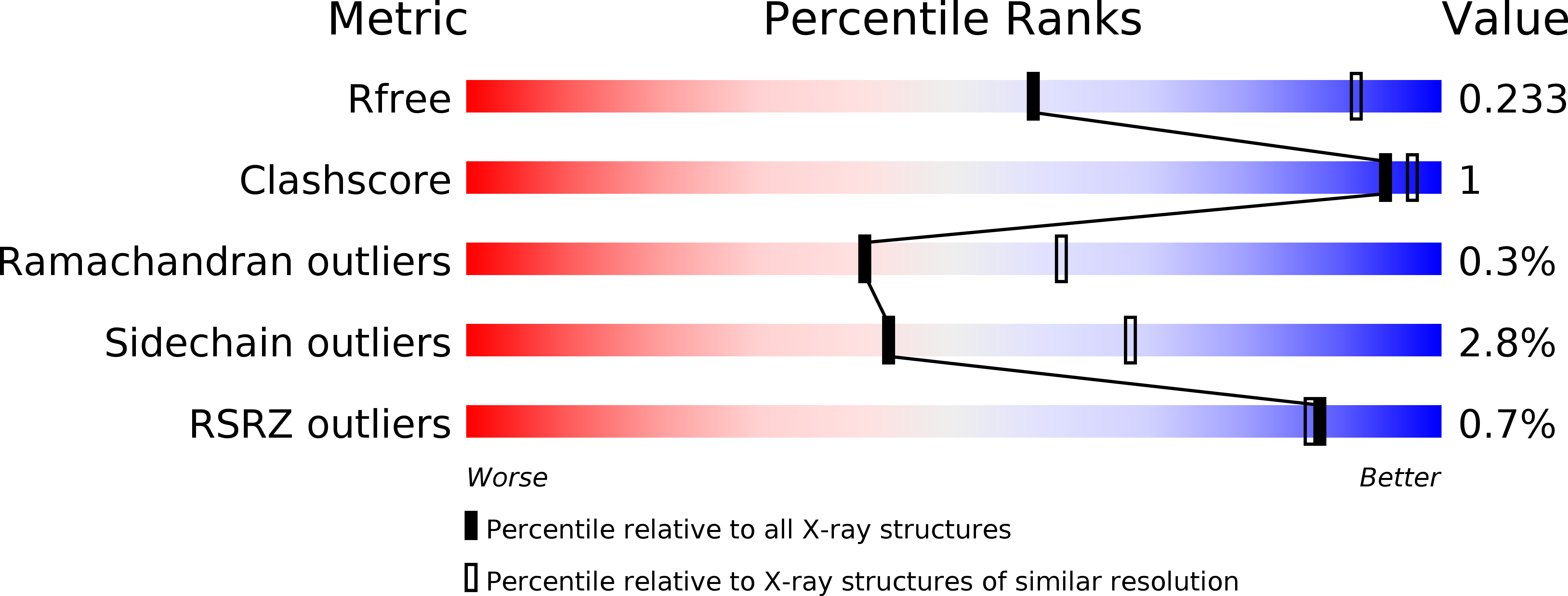
R-Value Free:
0.23
R-Value Work:
0.20
R-Value Observed:
0.20
Space Group:
P 43 21 2