3V3T image
Deposition Date 2011-12-14
Release Date 2012-05-16
Last Version Date 2024-02-28
Entry Detail
PDB ID:
3V3T
Title:
Crystal structure of Clostridium botulinum phage c-st TubZ
Biological Source:
Source Organism:
Host Organism:
Method Details:
Experimental Method:
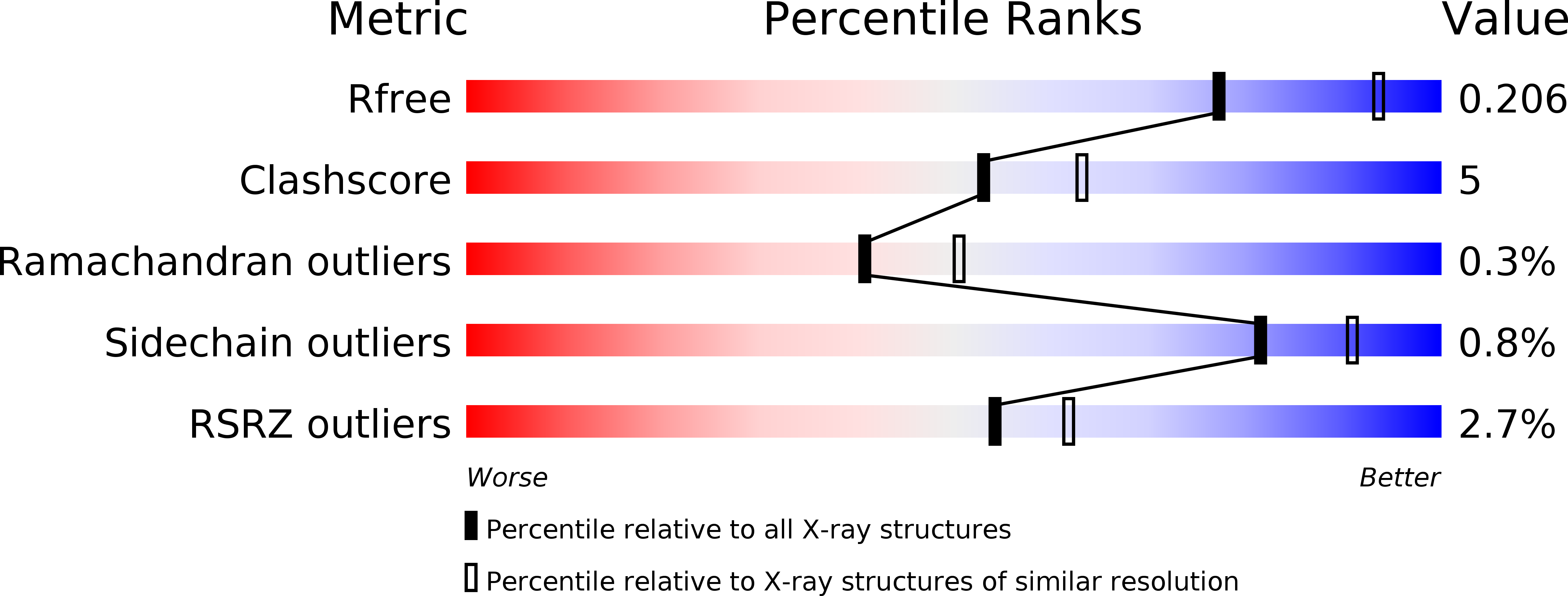
Resolution:
2.30 Å
R-Value Free:
0.21
R-Value Work:
0.16
R-Value Observed:
0.16
Space Group:
C 1 2 1
Macromolecular Entities
Polymer Type:polypeptide(L)
Molecule:Cell division GTPase FtsZ, diverged
Mutations:T100A
Chain IDs:A
Chain Length:360
Number of Molecules:1
Biological Source:Clostridium botulinum C
Primary Citation
Tubulin homolog TubZ in a phage-encoded partition system.
Proc.Natl.Acad.Sci.USA 109 7711 7716 (2012)
PMID: 22538818 DOI: 10.1073/pnas.1121546109

Abstact

Partition systems are responsible for the process whereby large and essential plasmids are accurately positioned to daughter cells during bacterial division. They are typically made of three components: a centromere-like DNA zone, an adaptor protein, and an assembling protein that is either a Walker-box ATPase (type I) or an actin-like ATPase (type II). A recently described type III segregation system has a tubulin/FtsZ-like protein, called TubZ, for plasmid movement. Here, we present the 2.3 Å structure and dynamic assembly of a TubZ tubulin homolog from a bacteriophage and unravel the Clostridium botulinum phage c-st type III partition system. Using biochemical and biophysical approaches, we prove that a gene upstream from tubZ encodes the partner TubR and localize the centromeric region (tubS), both of which are essential for anchoring phage DNA to the motile TubZ filaments. Finally, we describe a conserved fourth component, TubY, which modulates the TubZ-R-S complex interaction.

Legend

Protein

Chemical

Disease

Primary Citation of related structures