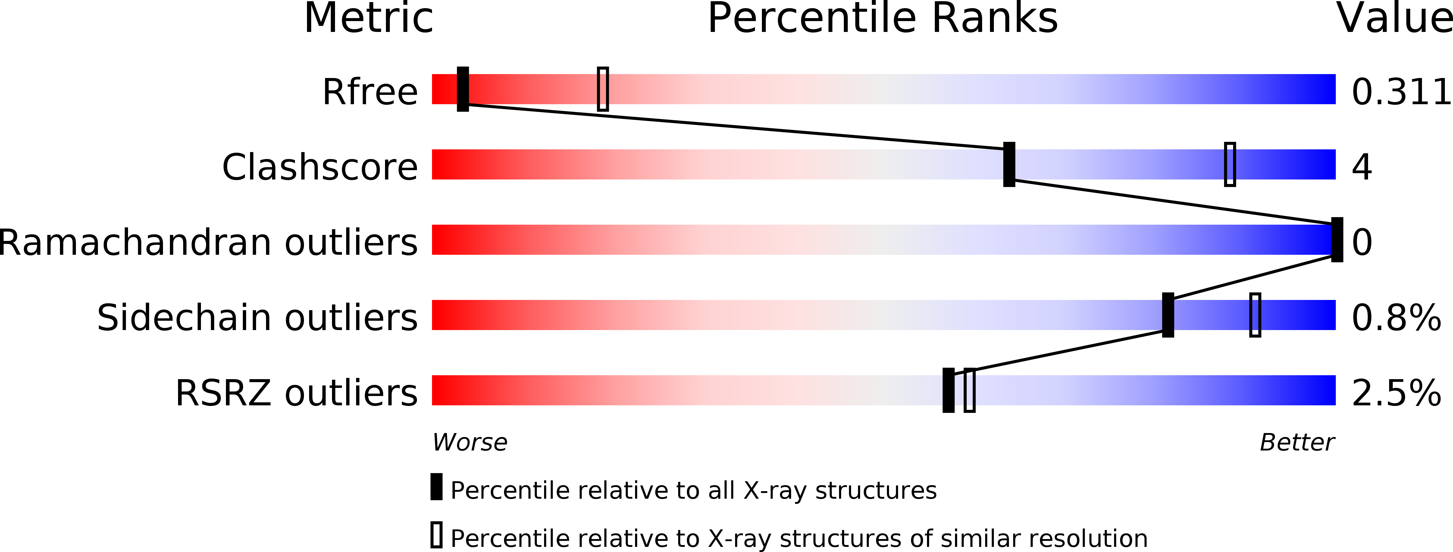
3V2W image
Entry Detail
PDB ID:
3V2W
Keywords:
Title:
Crystal Structure of a Lipid G protein-Coupled Receptor at 3.35A
Biological Source:
PDB Version:
Deposition Date:
2011-12-12
Release Date:
2012-02-15
Method Details:
Experimental Method:
Resolution:
3.35 Å
R-Value Free:
0.28
R-Value Work:
0.22
R-Value Observed:
0.22
Space Group:
P 21 21 2
Macromolecular Entities
Polymer Type:polypeptide(L)
Description:Sphingosine 1-phosphate receptor 1, Lysozyme chimera
Mutations:C1054T, C1097A
Chain IDs:A
Chain Length:520
Number of Molecules:1
Biological Source:Homo sapiens, Enterobacteria phage T4
Modified Residue
Compound ID Chain ID Parent Comp ID Details 2D Image
ASN A ASN GLYCOSYLATION SITE
Primary Citation
Crystal structure of a lipid G protein-coupled receptor.
Science 335 851 855 (2012)
PMID: 22344443 DOI: 10.1126/science.1215904

Abstact

The lyso-phospholipid sphingosine 1-phosphate modulates lymphocyte trafficking, endothelial development and integrity, heart rate, and vascular tone and maturation by activating G protein-coupled sphingosine 1-phosphate receptors. Here, we present the crystal structure of the sphingosine 1-phosphate receptor 1 fused to T4-lysozyme (S1P(1)-T4L) in complex with an antagonist sphingolipid mimic. Extracellular access to the binding pocket is occluded by the amino terminus and extracellular loops of the receptor. Access is gained by ligands entering laterally between helices I and VII within the transmembrane region of the receptor. This structure, along with mutagenesis, agonist structure-activity relationship data, and modeling, provides a detailed view of the molecular recognition and requirement for hydrophobic volume that activates S1P(1), resulting in the modulation of immune and stromal cell responses.

Legend

Protein

Chemical

Disease

Primary Citation of related structures