
Deposition Date
2011-12-09
Release Date
2012-01-11
Last Version Date
2023-09-13
Entry Detail
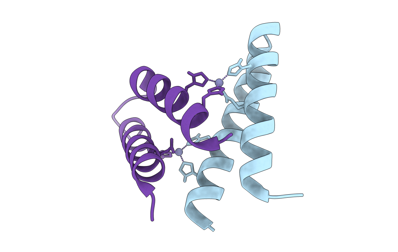
PDB ID:
3V1E
Keywords:
Title:
Crystal structure of de novo designed MID1-zinc H12E mutant
Biological Source:
Source Organism(s):
ARTIFICIAL GENE (Taxon ID: 32630)
Expression System(s):
Method Details:
Experimental Method:
Resolution:
1.07 Å
R-Value Free:
0.15
R-Value Work:
0.14
R-Value Observed:
0.14
Space Group:
P 21 21 21
