Deposition Date
2011-12-08
Release Date
2012-02-22
Last Version Date
2023-11-08
Entry Detail
PDB ID:
3V0S
Keywords:
Title:
Crystal Structure of Perakine Reductase, Founder Member of a Novel AKR Subfamily with Unique Conformational Changes during NADPH Binding
Biological Source:
Source Organism(s):
Rauvolfia serpentina (Taxon ID: 4060)
Expression System(s):
Method Details:
Experimental Method:
Resolution:
1.77 Å
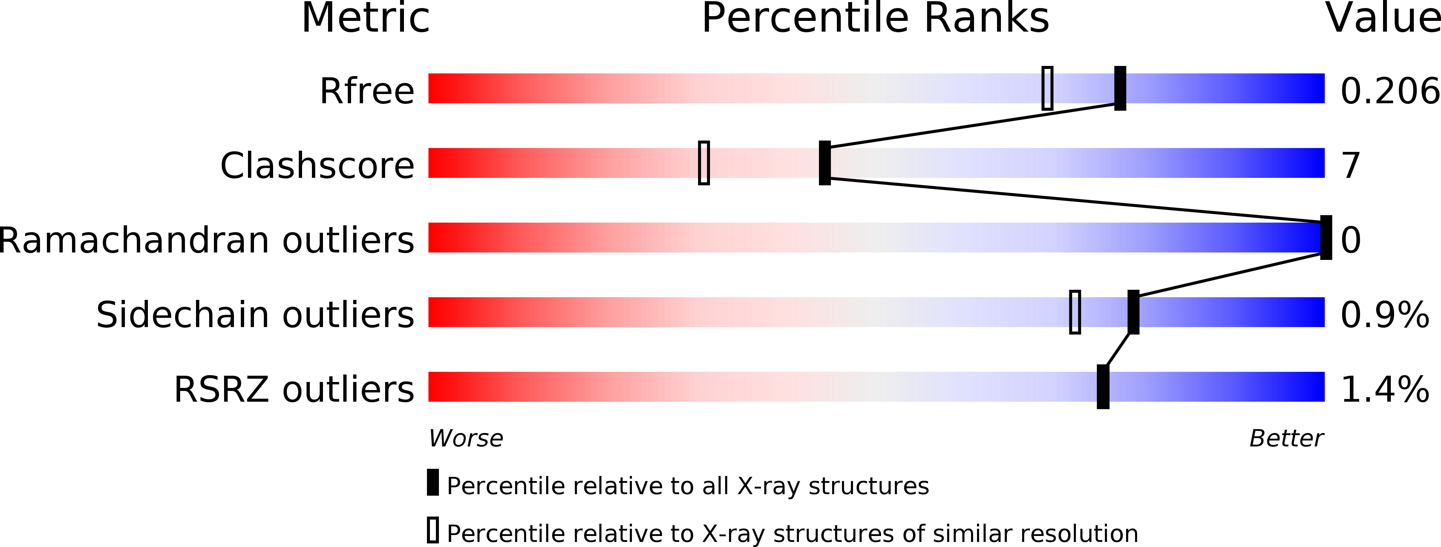
R-Value Free:
0.21
R-Value Work:
0.18
R-Value Observed:
0.18
Space Group:
P 32 2 1