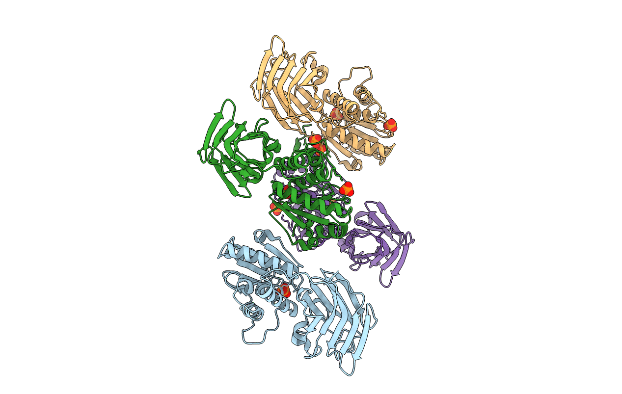
3V0G image
Entry Detail
PDB ID:
3V0G
Keywords:
Title:
Crystal structure of Ciona intestinalis voltage sensor-containing phosphatase (Ci-VSP), residues 241-576(C363S), form III
Biological Source:
Source Organism:
Host Organism:
PDB Version:
Deposition Date:
2011-12-08
Release Date:
2012-05-09
Method Details:
Experimental Method:
Resolution:
1.60 Å
R-Value Free:
0.29
R-Value Work:
0.25
R-Value Observed:
0.25
Space Group:
P 1
Macromolecular Entities
Polymer Type:polypeptide(L)
Description:Voltage-sensor containing phosphatase
Mutations:C363S
Chain IDs:A, B, C, D
Chain Length:339
Number of Molecules:4
Biological Source:Ciona intestinalis
Ligand Molecules
Primary Citation
A glutamate switch controls voltage-sensitive phosphatase function.
Nat.Struct.Mol.Biol. 19 633 641 (2012)
PMID: 22562138 DOI: 10.1038/nsmb.2289

Abstact

The Ciona intestinalis voltage-sensing phosphatase (Ci-VSP) couples a voltage-sensing domain (VSD) to a lipid phosphatase that is similar to the tumor suppressor PTEN. How the VSD controls enzyme function has been unclear. Here, we present high-resolution crystal structures of the Ci-VSP enzymatic domain that reveal conformational changes in a crucial loop, termed the 'gating loop', that controls access to the active site by a mechanism in which residue Glu411 directly competes with substrate. Structure-based mutations that restrict gating loop conformation impair catalytic function and demonstrate that Glu411 also contributes to substrate selectivity. Structure-guided mutations further define an interaction between the gating loop and linker that connects the phosphatase to the VSD for voltage control of enzyme activity. Together, the data suggest that functional coupling between the gating loop and the linker forms the heart of the regulatory mechanism that controls voltage-dependent enzyme activation.

Legend

Protein

Chemical

Disease

Primary Citation of related structures