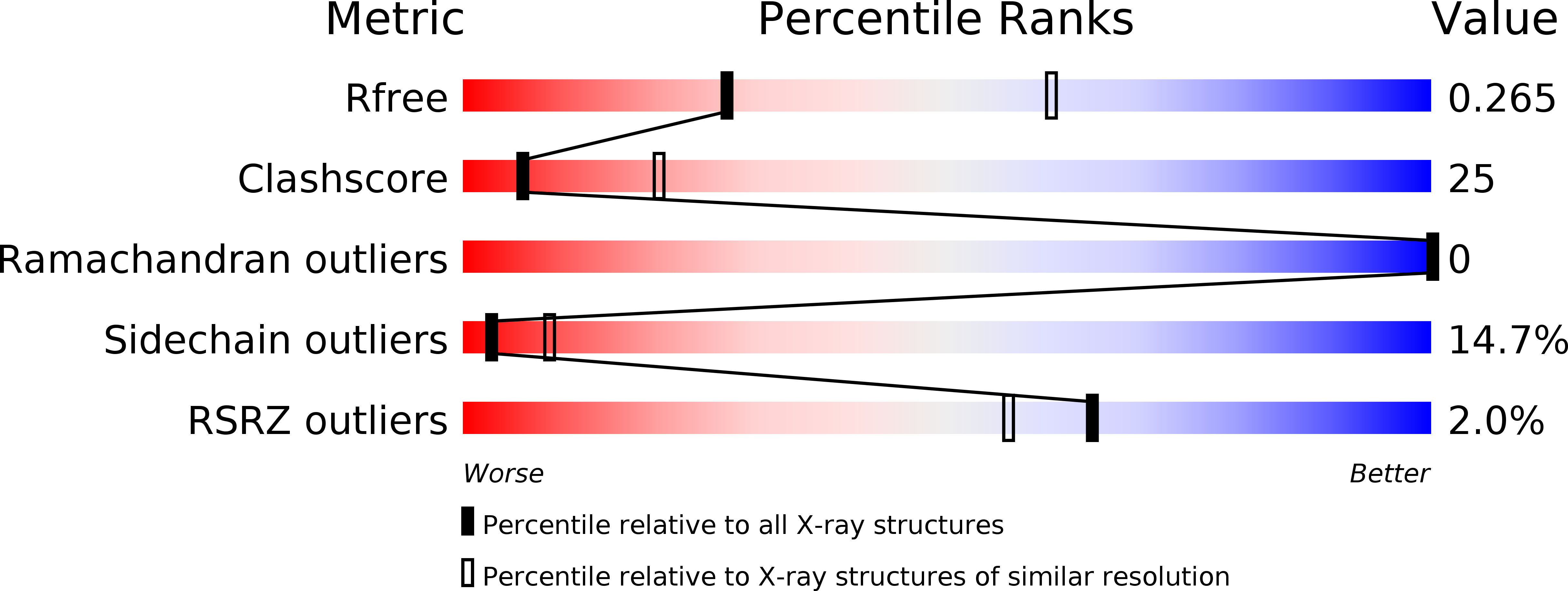
3UZ0 image
Deposition Date 2011-12-07
Release Date 2012-03-21
Last Version Date 2024-10-09
Entry Detail
PDB ID:
3UZ0
Title:
Crystal Structure of SpoIIIAH and SpoIIQ Complex
Biological Source:
Source Organism:
Host Organism:
Method Details:
Experimental Method:
Resolution:
2.82 Å
R-Value Free:
0.27
R-Value Work:
0.20
R-Value Observed:
0.21
Space Group:
P 21 21 21
Macromolecular Entities
Structures with similar UniProt ID
Protein Blast
Polymer Type:polypeptide(L)
Molecule:Stage III sporulation protein AH
Gene (Uniprot):spoIIIAH
Chain IDs:A, C
Chain Length:133
Number of Molecules:2
Biological Source:Bacillus subtilis
Structures with similar UniProt ID
Protein Blast
Polymer Type:polypeptide(L)
Molecule:Stage II sporulation protein Q
Gene (Uniprot):spoIIQ
Chain IDs:B, D
Chain Length:152
Number of Molecules:2
Biological Source:Bacillus subtilis
Modified Residue
Compound ID Chain ID Parent Comp ID Details 2D Image
MSE A MET SELENOMETHIONINE
Ligand Molecules
Primary Citation
Structure of the basal components of a bacterial transporter.
Proc.Natl.Acad.Sci.USA 109 5446 5451 (2012)
PMID: 22431613 DOI: 10.1073/pnas.1120113109

Abstact

Proteins SpoIIQ and SpoIIIAH interact through two membranes to connect the forespore and the mother cell during endospore development in the bacterium Bacillus subtilis. SpoIIIAH consists of a transmembrane segment and an extracellular domain with similarity to YscJ proteins. YscJ proteins form large multimeric rings that are the structural scaffolds for the assembly of type III secretion systems in gram-negative bacteria. The predicted ring-forming motif of SpoIIIAH and other evidence led to the model that SpoIIQ and SpoIIIAH form the core components of a channel or transporter through which the mother cell nurtures forespore development. Therefore, to understand the roles of SpoIIIAH and SpoIIQ in channel formation, it is critical to determine whether SpoIIIAH adopts a ring-forming structural motif, and whether interaction of SpoIIIAH with SpoIIQ would preclude ring formation. We report a 2.8-Å resolution structure of a complex of SpoIIQ and SpoIIIAH. SpoIIIAH folds into the ring-building structural motif, and modeling shows that the structure of the SpoIIQ-SpoIIIAH complex is compatible with forming a symmetrical oligomer that is similar to those in type III systems. The inner diameters of the two most likely ring models are large enough to accommodate several copies of other integral membrane proteins. SpoIIQ contains a LytM domain, which is found in metalloendopeptidases, but lacks residues important for metalloprotease activity. Other LytM domains appear to be involved in protein-protein interactions. We found that the LytM domain of SpoIIQ contains an accessory region that interacts with SpoIIIAH.

Legend

Protein

Chemical

Disease

Primary Citation of related structures