3UYO image
Deposition Date 2011-12-06
Release Date 2011-12-28
Last Version Date 2023-09-13
Entry Detail
PDB ID:
3UYO
Title:
Crystal structure of monobody SH13/ABL1 SH2 domain complex
Biological Source:
Source Organism:
Homo sapiens (Taxon ID: 9606)
Host Organism:
Method Details:
Experimental Method:
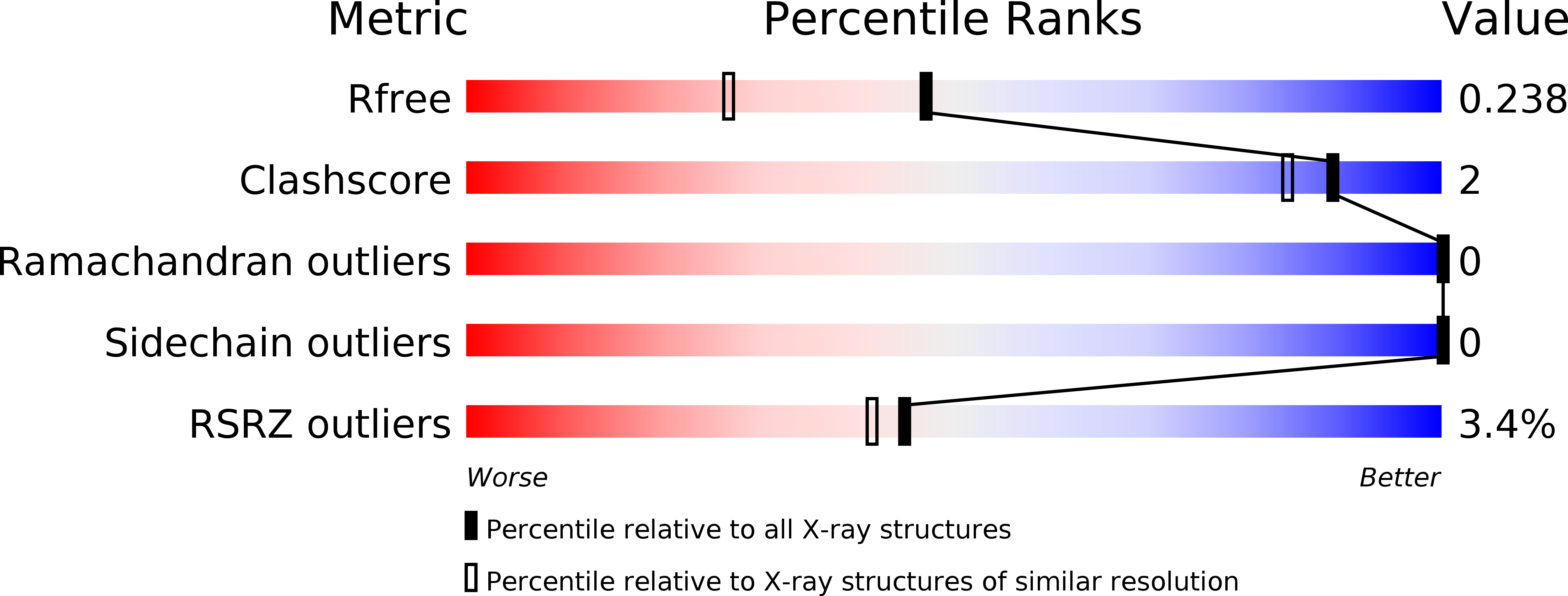
Resolution:
1.83 Å
R-Value Free:
0.23
R-Value Work:
0.18
R-Value Observed:
0.19
Space Group:
P 21 21 2
Macromolecular Entities
Polymer Type:polypeptide(L)
Molecule:Tyrosine-protein kinase ABL1
Gene (Uniprot):ABL1
Chain IDs:A
Chain Length:123
Number of Molecules:1
Biological Source:Homo sapiens
Polymer Type:polypeptide(L)
Molecule:Monobody SH13
Chain IDs:B (auth: D)
Chain Length:95
Number of Molecules:1
Biological Source:Homo sapiens
Ligand Molecules
Primary Citation
Teaching an old scaffold new tricks: monobodies constructed using alternative surfaces of the FN3 scaffold.
J.Mol.Biol. 415 393 405 (2012)
PMID: 22198408 DOI: 10.1016/j.jmb.2011.12.019

Abstact

The fibronectin type III domain (FN3) has become one of the most widely used non-antibody scaffolds for generating new binding proteins. Because of its structural homology to the immunoglobulin domain, combinatorial libraries of FN3 designed to date have primarily focused on introducing amino acid diversity into three loops that are equivalent to antibody complementarity-determining regions. Here, we report an FN3 library that utilizes alternative positions for presenting amino acid diversity. We diversified positions on a β-sheet and surface loops that together form a concave surface. The new library produced binding proteins (termed "monobodies") to multiple target proteins, generally with similar efficacy as the original, loop-focused library. The crystal structure of a monobody generated from the new library in complex with its target, the Abl SH2 domain, revealed that a concave surface of the monobody, as intended in our design, bound to a convex surface of the target with the interface area being among the largest of published structures of monobody-target complexes. This mode of interaction differs from a common binding mode for single-domain antibodies and antibody mimics in which recognition loops recognize clefts in targets. Together, this work illustrates the utilization of different surfaces of a single immunoglobulin-like scaffold to generate binding proteins with distinct characteristics.

Legend

Protein

Chemical

Disease

Primary Citation of related structures