3UYK image
Entry Detail
PDB ID:
3UYK
Keywords:
Title:
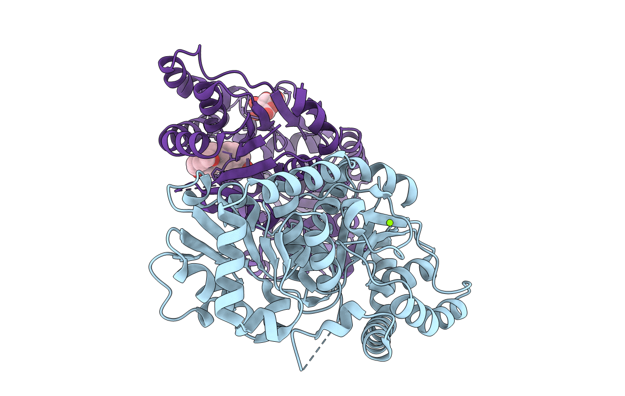
Spinosyn Rhamnosyltransferase SpnG complexed with spinosyn aglycone
Biological Source:
Source Organism:
Host Organism:
PDB Version:
Deposition Date:
2011-12-06
Release Date:
2012-02-15
Method Details:
Experimental Method:
Resolution:
1.70 Å
R-Value Free:
0.23
R-Value Work:
0.18
R-Value Observed:
0.19
Space Group:
P 1
Macromolecular Entities
Polymer Type:polypeptide(L)
Description:NDP-rhamnosyltransferase
Chain IDs:A, B
Chain Length:387
Number of Molecules:2
Biological Source:Saccharopolyspora spinosa
Primary Citation
Structural Studies of the Spinosyn Rhamnosyltransferase, SpnG.
Biochemistry 51 1213 1222 (2012)
PMID: 22283226 DOI: 10.1021/bi201860q

Abstact

Spinosyns A and D (spinosad), like many other complex polyketides, are tailored near the end of their biosyntheses through the addition of sugars. SpnG, which catalyzes their 9-OH rhamnosylation, is also capable of adding other monosaccharides to the spinosyn aglycone (AGL) from TDP-sugars; however, the substitution of UDP-D-glucose for TDP-D-glucose as the donor substrate is known to result in a >60000-fold reduction in k(cat). Here, we report the structure of SpnG at 1.65 Å resolution, SpnG bound to TDP at 1.86 Å resolution, and SpnG bound to AGL at 1.70 Å resolution. The SpnG-TDP complex reveals how SpnG employs N202 to discriminate between TDP- and UDP-sugars. A conformational change of several residues in the active site is promoted by the binding of TDP. The SpnG-AGL complex shows that the binding of AGL is mediated via hydrophobic interactions and that H13, the potential catalytic base, is within 3 Å of the nucleophilic 9-OH group of AGL. A model for the Michaelis complex was constructed to reveal the features that allow SpnG to transfer diverse sugars; it also revealed that the rhamnosyl moiety is in a skew-boat conformation during the transfer reaction.

Legend

Protein

Chemical

Disease

Primary Citation of related structures