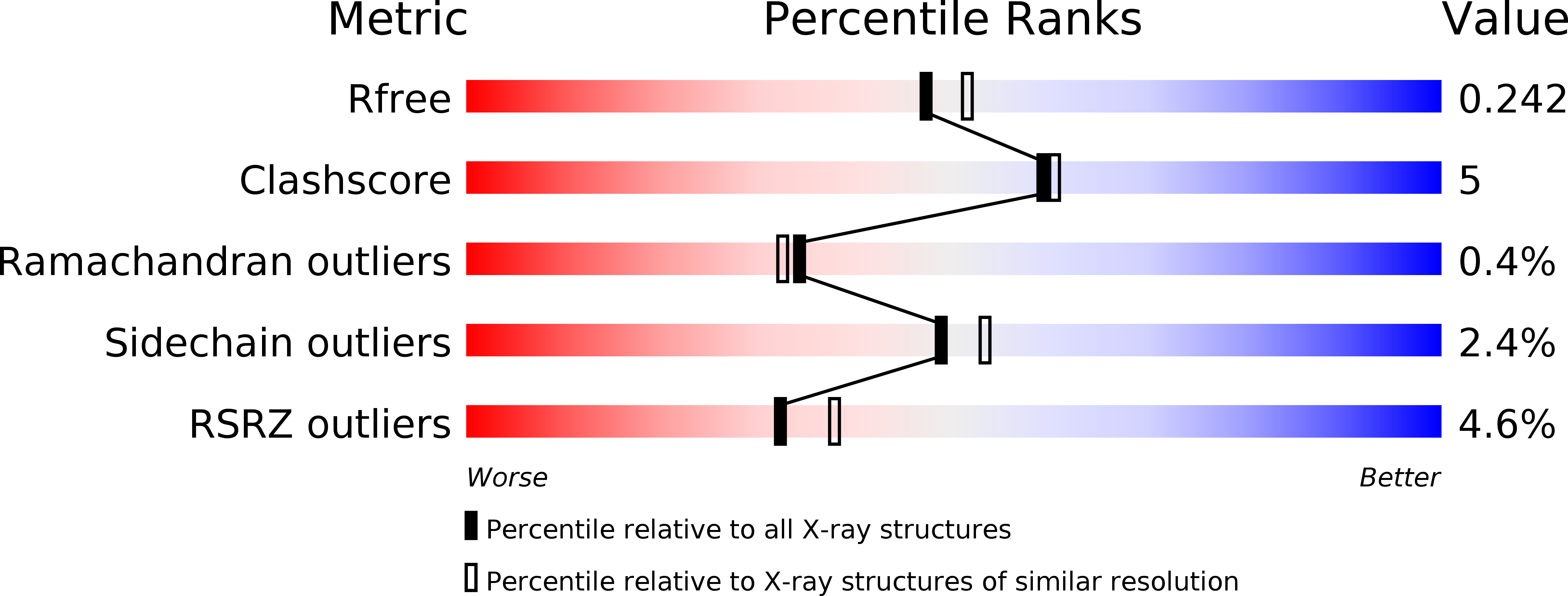
3UX8 image
Deposition Date 2011-12-04
Release Date 2012-02-08
Last Version Date 2024-02-28
Entry Detail
PDB ID:
3UX8
Title:
Crystal structure of UvrA
Biological Source:
Source Organism:
Geobacillus (Taxon ID: 550542)
Host Organism:
Method Details:
Experimental Method:
Resolution:
2.10 Å
R-Value Free:
0.24
R-Value Work:
0.21
R-Value Observed:
0.21
Space Group:
C 1 2 1
Macromolecular Entities
Polymer Type:polypeptide(L)
Molecule:Excinuclease ABC, A subunit
Chain IDs:A
Chain Length:670
Number of Molecules:1
Biological Source:Geobacillus
Primary Citation
Structure and mechanism of the UvrA-UvrB DNA damage sensor.
Nat.Struct.Mol.Biol. 19 291 298 (2012)
PMID: 22307053 DOI: 10.1038/nsmb.2240

Abstact

Nucleotide excision repair (NER) is used by all organisms to eliminate DNA lesions. We determined the structure of the Geobacillus stearothermophilus UvrA-UvrB complex, the damage-sensor in bacterial NER and a new structure of UvrA. We observe that the DNA binding surface of UvrA, previously found in an open shape that binds damaged DNA, also exists in a closed groove shape compatible with native DNA only. The sensor contains two UvrB molecules that flank the UvrA dimer along the predicted path for DNA, ~80 Å from the lesion. We show that the conserved signature domain II of UvrA mediates a nexus of contacts among UvrA, UvrB and DNA. Further, in our new structure of UvrA, this domain adopts an altered conformation while an adjacent nucleotide binding site is vacant. Our findings raise unanticipated questions about NER and also suggest a revised picture of its early stages.

Legend

Protein

Chemical

Disease

Primary Citation of related structures