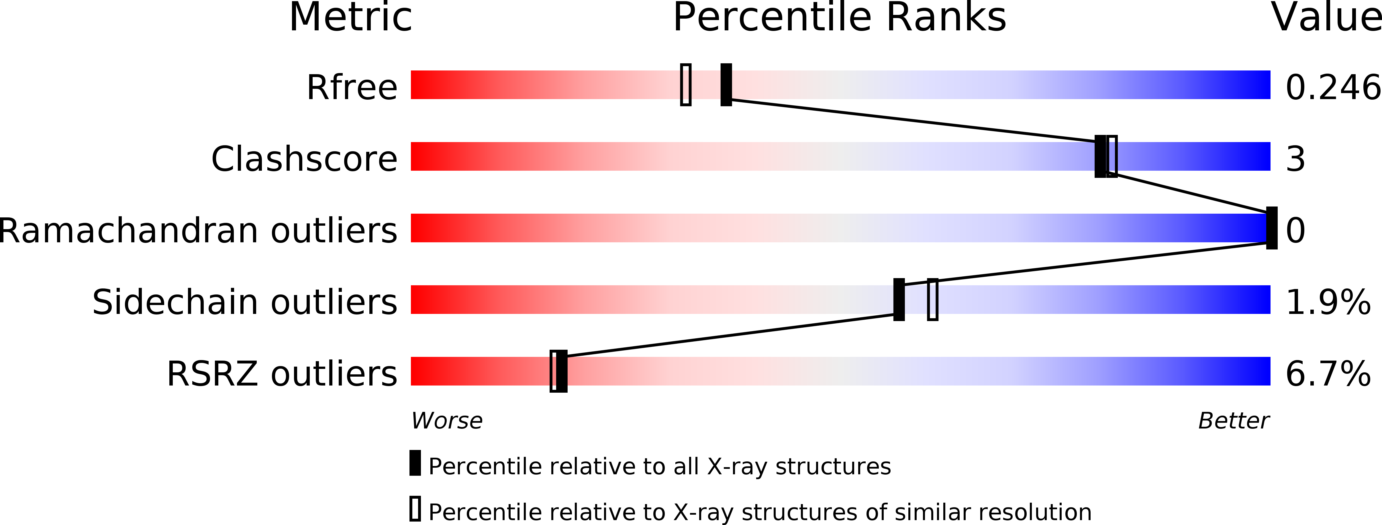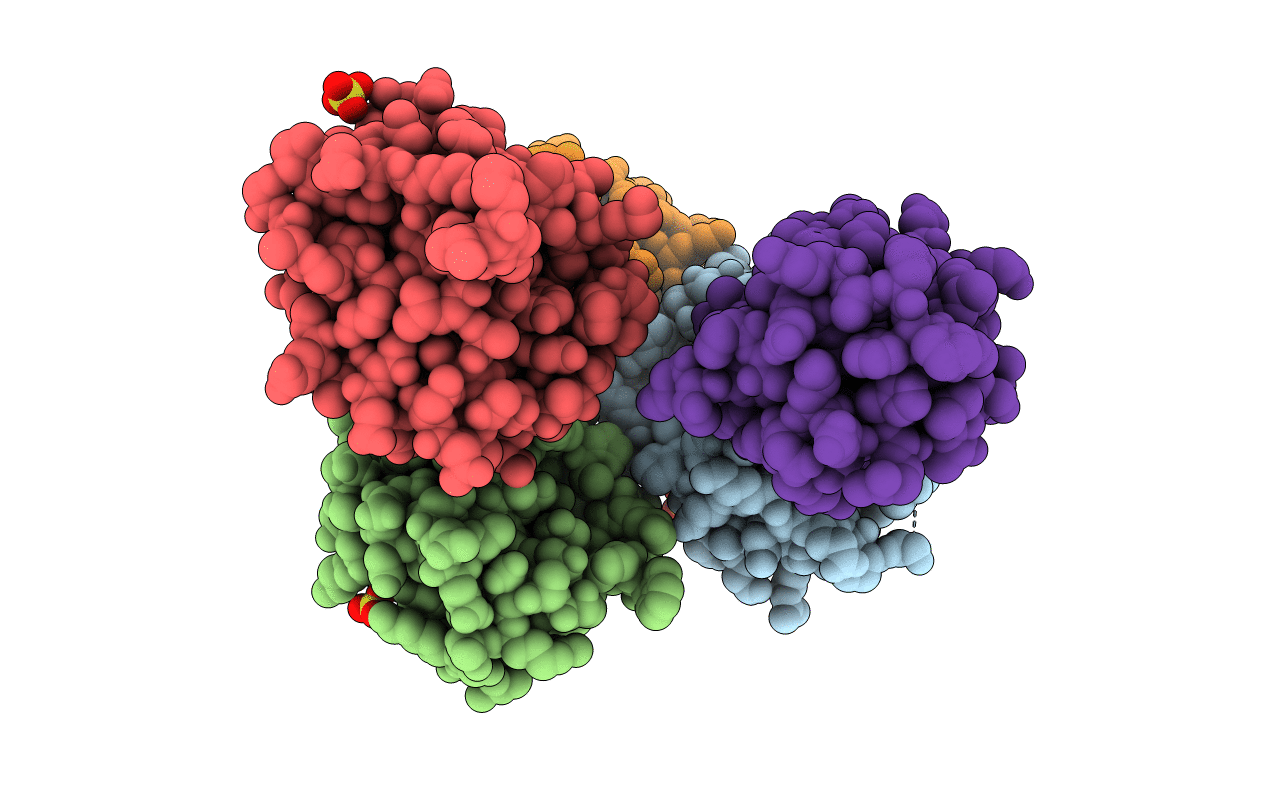
3UVT image
Deposition Date 2011-11-30
Release Date 2012-04-11
Last Version Date 2024-11-20
Entry Detail
PDB ID:
3UVT
Keywords:
Title:
Crystal structure of the third catalytic domain of ERp46
Biological Source:
Source Organism:
Homo sapiens (Taxon ID: 9606)
Host Organism:
Method Details:
Experimental Method:
Resolution:
2.00 Å
R-Value Free:
0.24
R-Value Work:
0.20
R-Value Observed:
0.20
Space Group:
P 1 21 1
Macromolecular Entities
Polymer Type:polypeptide(L)
Molecule:Thioredoxin domain-containing protein 5
Gene (Uniprot):TXNDC5
Chain IDs:A, B, C, D, E
Chain Length:111
Number of Molecules:5
Biological Source:Homo sapiens
Ligand Molecules
Primary Citation
Structure of the third catalytic domain of the protein disulfide isomerase ERp46.
Acta Crystallogr.,Sect.F 68 378 381 (2012)
PMID: 22505402 DOI: 10.1107/S1744309112005866

Abstact

Protein disulfide isomerases are responsible for catalyzing the proper oxidation and isomerization of disulfide bonds of newly synthesized proteins in the endoplasmic reticulum. Here, the crystal structure of the third catalytic domain of protein disulfide isomerase ERp46 (also known as protein disulfide isomerase A5 and TXNDC5) was determined to 2.0 Å resolution. The structure shows a typical thioredoxin-like fold, but also identifies regions of high structural variability. In particular, the loop between helix α2 and strand β3 adopts strikingly different conformations among the five chains of the asymmetric unit. Cys381 and Cys388 form a structural disulfide and its absence in one of the molecules leads to dramatic conformational changes. The tryptophan residue Trp349 of this molecule inserts into the cavity formed by helices α1 and α3 of a neighbouring molecule, potentially mimicking the interactions of ERp46 with misfolded substrates.

Legend

Protein

Chemical

Disease

Primary Citation of related structures