Deposition Date
2011-11-28
Release Date
2012-09-19
Last Version Date
2024-11-13
Entry Detail
PDB ID:
3UUN
Keywords:
Title:
Crystal Structure of N-terminal first spectrin repeat of dystrophin
Biological Source:
Source Organism(s):
Homo sapiens (Taxon ID: 9606)
Expression System(s):
Method Details:
Experimental Method:
Resolution:
2.30 Å
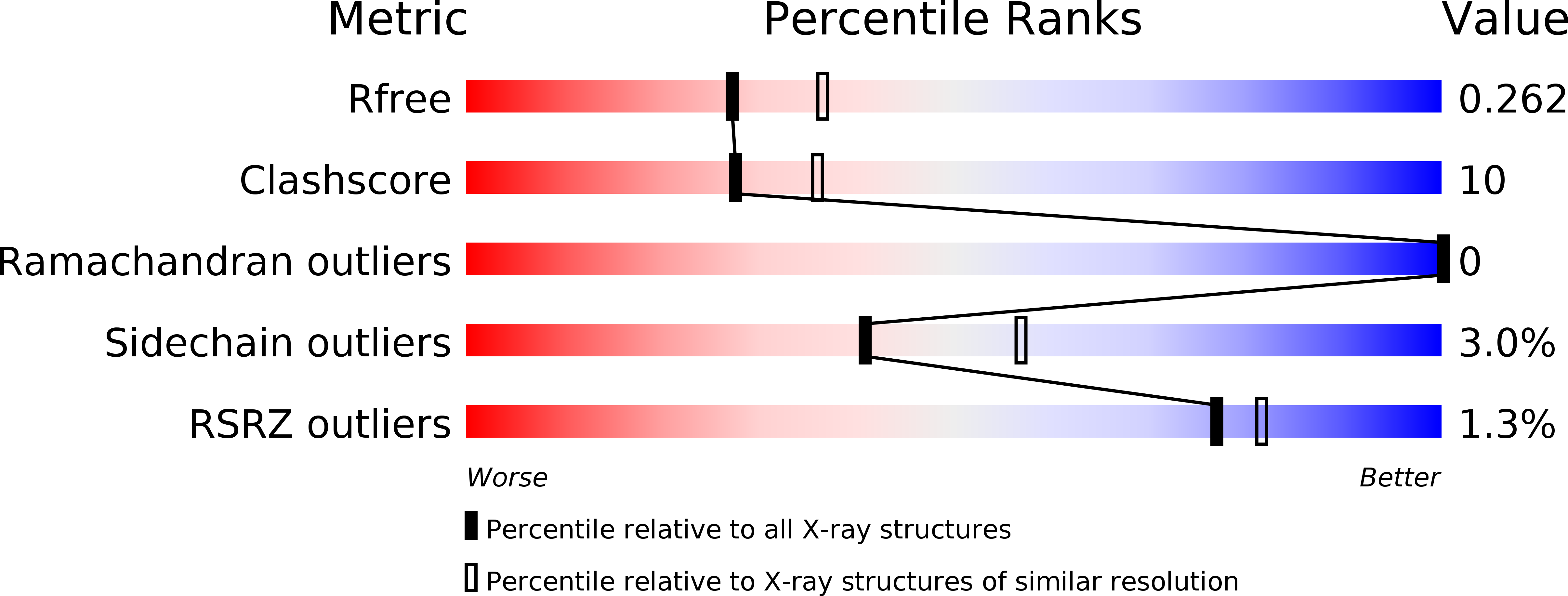
R-Value Free:
0.25
R-Value Work:
0.18
R-Value Observed:
0.19
Space Group:
P 32 2 1