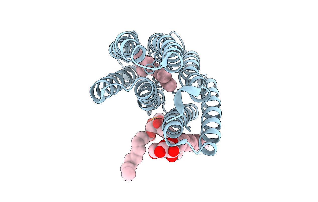
3UTW image
Entry Detail
PDB ID:
3UTW
Title:
Crystal structure of bacteriorhodopsin mutant P50A/Y57F
Biological Source:
Source Organism:
PDB Version:
Deposition Date:
2011-11-27
Release Date:
2012-05-09
Method Details:
Experimental Method:
Resolution:
2.40 Å
R-Value Free:
0.24
R-Value Work:
0.20
R-Value Observed:
0.20
Space Group:
C 2 2 21
Macromolecular Entities
Polymer Type:polypeptide(L)
Description:Bacteriorhodopsin
Mutations:P50A, Y57F
Chain IDs:A
Chain Length:249
Number of Molecules:1
Biological Source:Halobacterium sp.
Primary Citation
Shifting hydrogen bonds may produce flexible transmembrane helices.
Proc.Natl.Acad.Sci.USA 109 8121 8126 (2012)
PMID: 22566663 DOI: 10.1073/pnas.1201298109

Abstact

The intricate functions of membrane proteins would not be possible without bends or breaks that are remarkably common in transmembrane helices. The frequent helix distortions are nevertheless surprising because backbone hydrogen bonds should be strong in an apolar membrane, potentially rigidifying helices. It is therefore mysterious how distortions can be generated by the evolutionary currency of random point mutations. Here we show that we can engineer a transition between distinct distorted helix conformations in bacteriorhodopsin with a single-point mutation. Moreover, we estimate the energetic cost of the conformational transitions to be smaller than 1 kcal/mol. We propose that the low energy of distortion is explained in part by the shifting of backbone hydrogen bonding partners. Consistent with this view, extensive backbone hydrogen bond shifts occur during helix conformational changes that accompany functional cycles. Our results explain how evolution has been able to liberally exploit transmembrane helix bending for the optimization of membrane protein structure, function, and dynamics.

Legend

Protein

Chemical

Disease

Primary Citation of related structures