3UT6 image
Deposition Date 2011-11-25
Release Date 2012-05-23
Last Version Date 2024-03-20
Entry Detail
PDB ID:
3UT6
Keywords:
Title:
Crystal structure of E. Coli PNP complexed with PO4 and formycin A
Biological Source:
Source Organism:
Method Details:
Experimental Method:
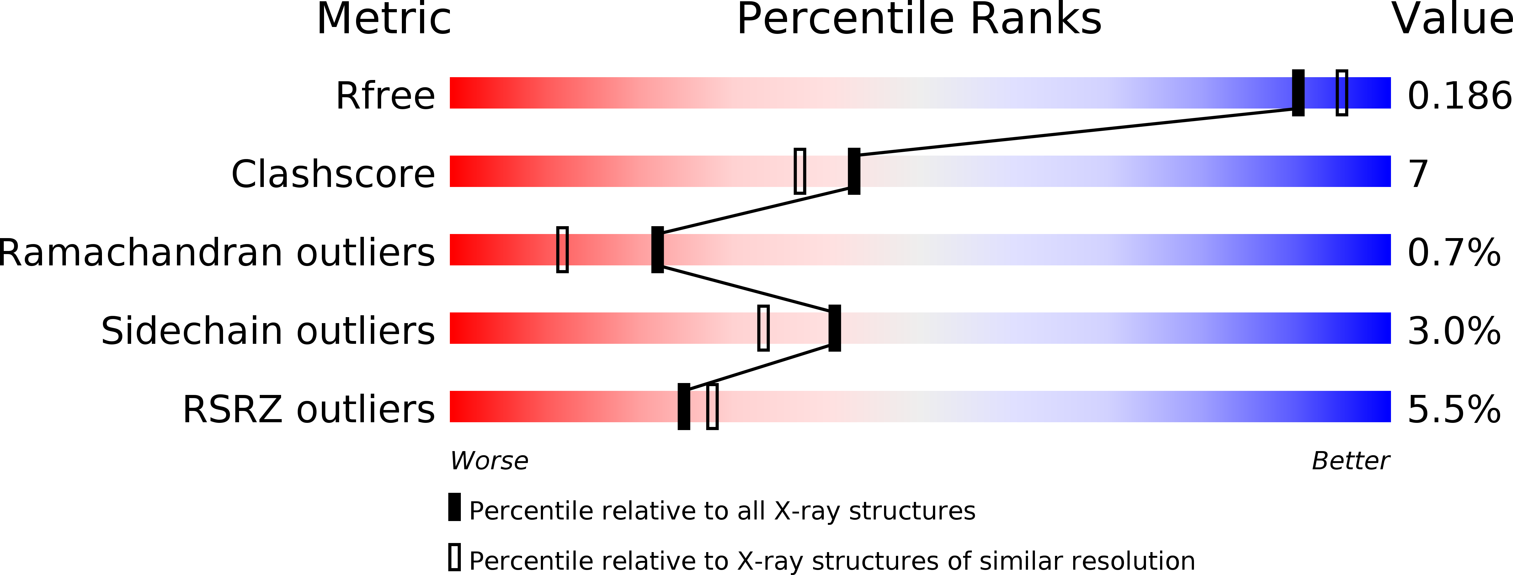
Resolution:
1.90 Å
R-Value Free:
0.18
R-Value Work:
0.16
R-Value Observed:
0.16
Space Group:
P 61 2 2
Macromolecular Entities
Polymer Type:polypeptide(L)
Molecule:Purine nucleoside phosphorylase deoD-type
Gene (Uniprot):deoD
Chain IDs:A, B, C
Chain Length:237
Number of Molecules:3
Biological Source:Escherichia coli
Primary Citation
New phosphate binding sites in the crystal structure of Escherichia coli purine nucleoside phosphorylase complexed with phosphate and formycin A.
Febs Lett. 586 967 971 (2012)
PMID: 22569248 DOI: 10.1016/j.febslet.2012.02.039

Abstact

Purine nucleoside phosphorylase (PNP) from Escherichia coli is a homohexamer that catalyses the phosphorolytic cleavage of the glycosidic bond of purine nucleosides. The first crystal structure of the ternary complex of this enzyme (with a phosphate ion and formycin A), which is biased by neither the presence of an inhibitor nor sulfate as a precipitant, is presented. The structure reveals, in some active sites, an unexpected and never before observed binding site for phosphate and exhibits a stoichiometry of two phosphate molecules per enzyme subunit. Moreover, in these active sites, the phosphate and nucleoside molecules are found not to be in direct contact. Rather, they are bridged by three water molecules that occupy the "standard" phosphate binding site.

Legend

Protein

Chemical

Disease

Primary Citation of related structures