3USY image
Deposition Date 2011-11-24
Release Date 2011-12-14
Last Version Date 2023-11-08
Entry Detail
PDB ID:
3USY
Keywords:
Title:
Crystal structure of Flig (residue 116-343) from H. Pylori
Biological Source:
Source Organism:
Host Organism:
Method Details:
Experimental Method:
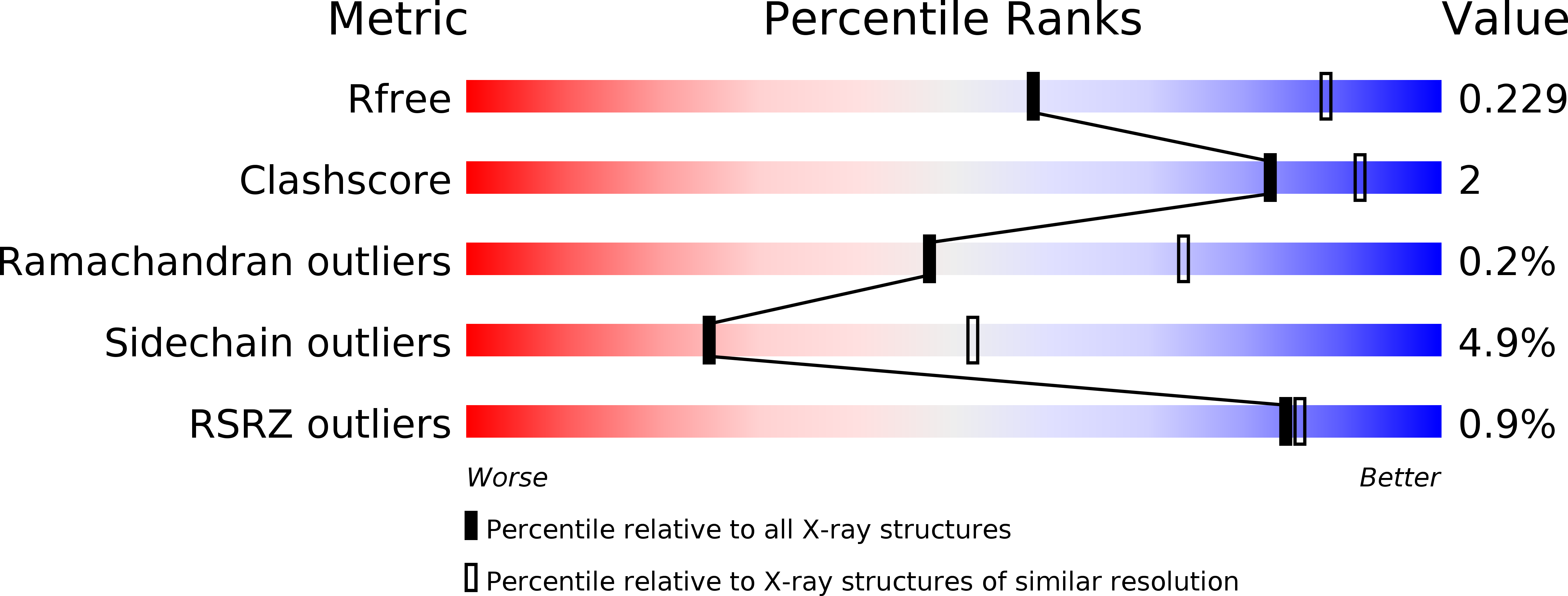
Resolution:
2.71 Å
R-Value Free:
0.23
R-Value Work:
0.19
R-Value Observed:
0.19
Space Group:
C 1 2 1
Macromolecular Entities
Structures with similar UniProt ID
Protein Blast
Polymer Type:polypeptide(L)
Molecule:Flagellar motor switch protein
Gene (Uniprot):fliG
Chain IDs:A, B
Chain Length:234
Number of Molecules:2
Biological Source:Helicobacter pylori
Primary Citation
Multiple conformations of the FliG C-terminal domain provide insight into flagellar motor switching
Structure 20 315 325 (2012)
PMID: 22325779 DOI: 10.1016/j.str.2011.11.020

Abstact

Bacterial flagellar switching between counterclockwise and clockwise directions is mediated by the coupling of the chemotactic system and the motor switch complex. The conformational changes of FliG are closely associated with this switching mechanism. We present two crystal structures of FliG(MC) from Helicobacter pylori, each showing distinct domain orientations from previously solved structures. A 180° rotation of the charged ridge-containing C-terminal subdomain FliG(Cα1-6) that is prompted by the rotational freedom of Met245 psi and Phe246 phi at the MFXF motif was revealed. Studies on the swarming and swimming behavior of Escherichia coli mutants further identified the importance of the ₂₄₅MFXF₂₄₈ motif and a highly conserved residue, Asn216, in motor switching. Additionally, multiple conformations of FliG(Cα1-6) were demonstrated by intramolecular cysteine crosslinking. The conformational flexibility of FliGc leads us to propose a model that accounts for the symmetrical torque generation process and for the dynamics of the motor.

Legend

Protein

Chemical

Disease

Primary Citation of related structures