Deposition Date
2011-11-23
Release Date
2011-12-14
Last Version Date
2024-10-30
Entry Detail
PDB ID:
3USH
Keywords:
Title:
Crystal Structure of the Q2S0R5 protein from Salinibacter ruber, Northeast Structural Genomics Consortium Target SrR207
Biological Source:
Source Organism(s):
Salinibacter ruber (Taxon ID: 309807)
Expression System(s):
Method Details:
Experimental Method:
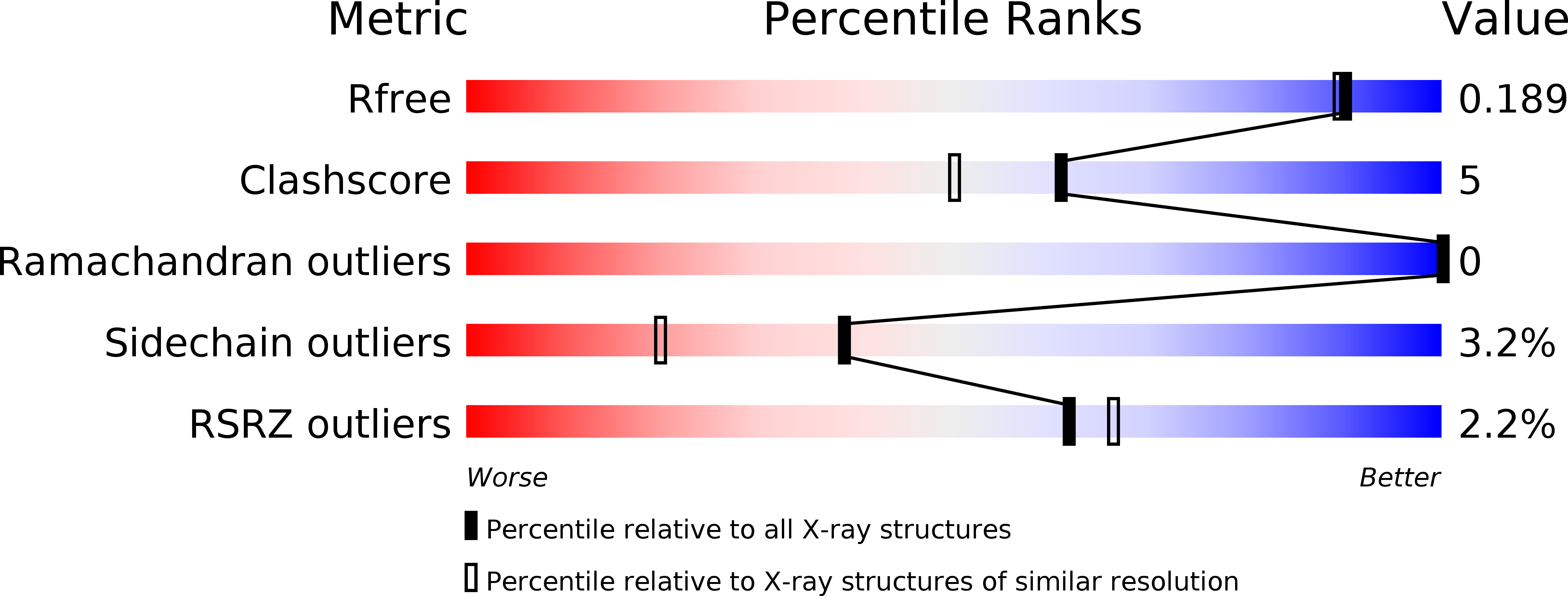
Resolution:
1.69 Å
R-Value Free:
0.19
R-Value Work:
0.17
R-Value Observed:
0.17
Space Group:
P 1 21 1