
Deposition Date
2011-11-22
Release Date
2012-03-28
Last Version Date
2024-11-06
Entry Detail
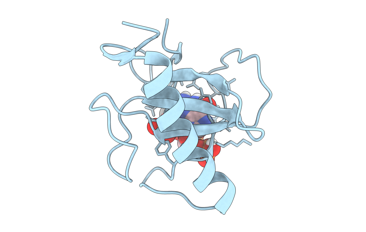
PDB ID:
3URP
Keywords:
Title:
Re-refinement of PDB entry 5RNT - ribonuclease T1 with guanosine-3',5'-diphosphate and phosphate ion bound
Biological Source:
Source Organism(s):
Aspergillus oryzae (Taxon ID: 5062)
Method Details:
Experimental Method:
Resolution:
3.19 Å
R-Value Free:
0.23
R-Value Work:
0.20
R-Value Observed:
0.20
Space Group:
I 2 3
