
Deposition Date
2011-11-16
Release Date
2012-02-01
Last Version Date
2024-11-06
Entry Detail
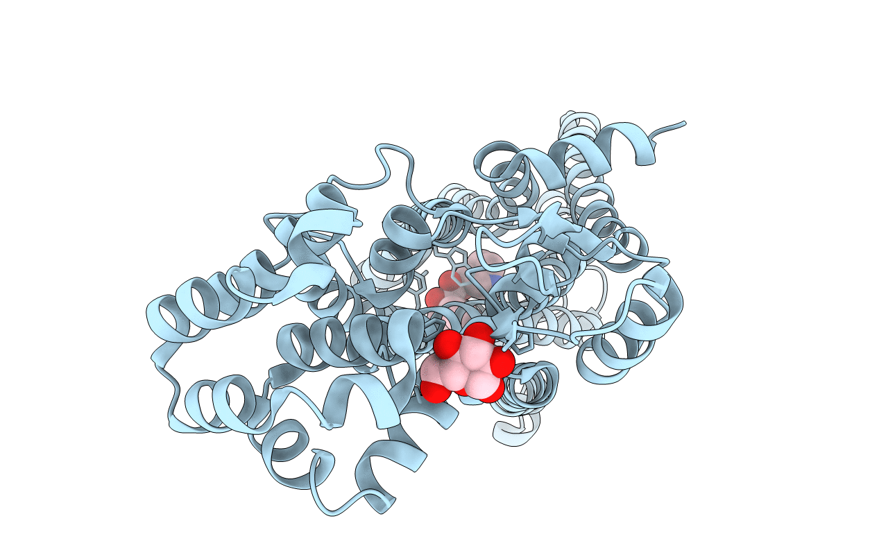
PDB ID:
3UON
Keywords:
Title:
Structure of the human M2 muscarinic acetylcholine receptor bound to an antagonist
Biological Source:
Source Organism(s):
Homo sapiens (Taxon ID: 9606)
Enterobacteria phage T4 (Taxon ID: 10665)
Enterobacteria phage T4 (Taxon ID: 10665)
Expression System(s):
Method Details:
Experimental Method:
Resolution:
3.00 Å
R-Value Free:
0.27
R-Value Work:
0.22
R-Value Observed:
0.22
Space Group:
P 1 21 1
