3UOA image
Entry Detail
PDB ID:
3UOA
Title:
Crystal structure of the MALT1 paracaspase (P21 form)
Biological Source:
Source Organism:
Host Organism:
PDB Version:
Deposition Date:
2011-11-16
Release Date:
2011-12-21
Method Details:
Experimental Method:
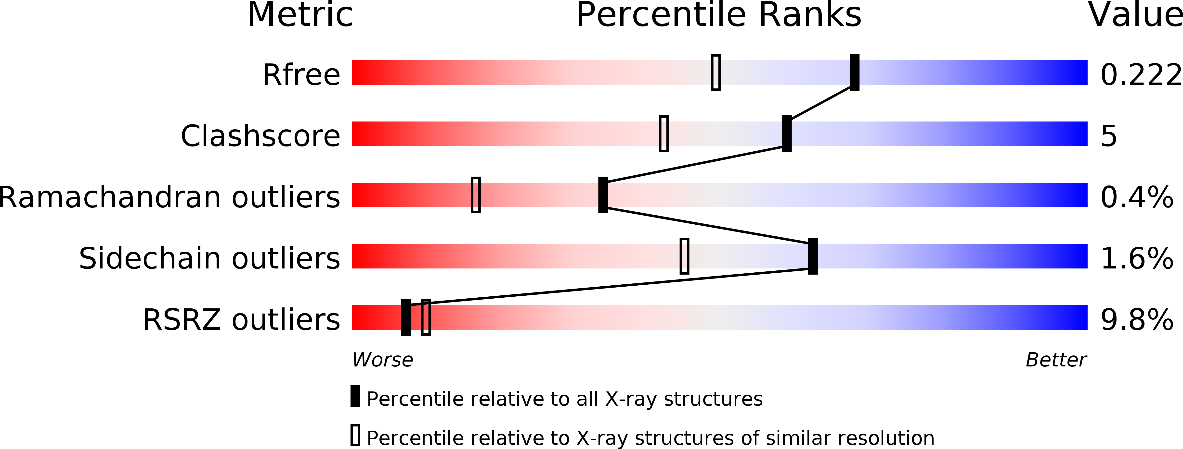
Resolution:
1.75 Å
R-Value Free:
0.22
R-Value Work:
0.19
R-Value Observed:
0.19
Space Group:
P 1 21 1
Macromolecular Entities
Polymer Type:polypeptide(L)
Description:Mucosa-associated lymphoid tissue lymphoma translocation protein 1
Chain IDs:A (auth: B), B (auth: C)
Chain Length:390
Number of Molecules:2
Biological Source:Homo sapiens
Polymer Type:polypeptide(L)
Description:Z-Val-Arg-Pro-DL-Arg-fluoromethylketone
Chain IDs:C (auth: L), D (auth: M)
Chain Length:6
Number of Molecules:2
Biological Source:
Ligand Molecules
Peptide-like Molecules
PRD_001076
Primary Citation
Crystal structure of the mucosa-associated lymphoid tissue lymphoma translocation 1 (MALT1) paracaspase region.
Proc.Natl.Acad.Sci.USA 108 21004 21009 (2011)
PMID: 22158899 DOI: 10.1073/pnas.1111708108

Abstact

The mucosa-associated lymphoid tissue lymphoma translocation 1 (MALT1) paracaspase, a key component of the Carma1/Bcl10/MALT1 signalosome, is critical for NF-κB signaling in multiple contexts. MALT1 is thought to function as a scaffold and protease to promote signaling; however, the biochemical and structural basis of paracaspase action remains largely unknown. Here we report the 1.75-Å resolution crystal structure of the MALT1 paracaspase region, which contains the paracaspase domain and an ensuing Ig-like domain. The paracaspase and the Ig domains appear as a single folding unit and interact with each other through extensive van der Waals contacts and hydrogen bonds. The paracaspase domain adopts a fold that is nearly identical to that of classic caspases and homodimerizes similarly to form an active protease. Unlike caspases, the active and mature form of the paracaspase domain remains a single uncleaved polypeptide and specifically recognizes the bound peptide inhibitor Val-Arg-Pro-Arg. In particular, the carboxyl-terminal amino acid Arg of the inhibitor is coordinated by three highly conserved acidic residues. This structure serves as an important framework for deciphering the function and mechanism of paracaspases exemplified by MALT1.

Legend

Protein

Chemical

Disease

Primary Citation of related structures