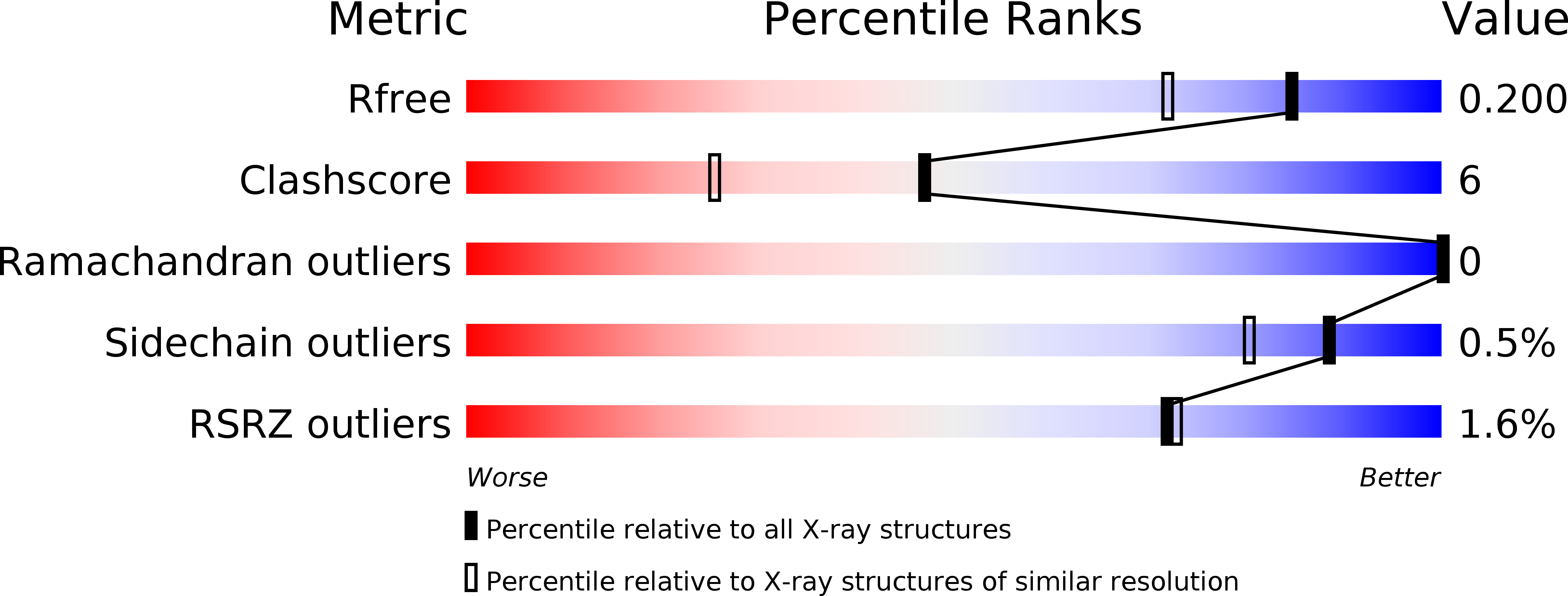
3UO1 image
Deposition Date 2011-11-16
Release Date 2012-07-25
Last Version Date 2024-11-27
Entry Detail
PDB ID:
3UO1
Keywords:
Title:
Structure of a monoclonal antibody complexed with its MHC-I antigen
Biological Source:
Source Organism:
Mus musculus (Taxon ID: 10090)
Method Details:
Experimental Method:
Resolution:
1.64 Å
R-Value Free:
0.20
R-Value Work:
0.18
R-Value Observed:
0.18
Space Group:
C 1 2 1
Macromolecular Entities
Polymer Type:polypeptide(L)
Molecule:ANTI-MHC-I MONOCLONAL ANTIBODY, 64-3-7 H CHAIN
Chain IDs:A (auth: H)
Chain Length:216
Number of Molecules:1
Biological Source:Mus musculus
Polymer Type:polypeptide(L)
Molecule:ANTI-MHC-I MONOCLONAL ANTIBODY, 64-3-7 L CHAIN
Chain IDs:B (auth: L)
Chain Length:218
Number of Molecules:1
Biological Source:Mus musculus
Polymer Type:polypeptide(L)
Molecule:H-2 class I histocompatibility antigen, L-D alpha chain
Gene (Uniprot):H2-L
Chain IDs:C (auth: P)
Chain Length:9
Number of Molecules:1
Biological Source:Mus musculus
Ligand Molecules
Primary Citation
The Peptide-receptive transition state of MHC class I molecules: insight from structure and molecular dynamics.
J.Immunol. 189 1391 1399 (2012)
PMID: 22753930 DOI: 10.4049/jimmunol.1200831

Abstact

MHC class I (MHC-I) proteins of the adaptive immune system require antigenic peptides for maintenance of mature conformation and immune function via specific recognition by MHC-I-restricted CD8(+) T lymphocytes. New MHC-I molecules in the endoplasmic reticulum are held by chaperones in a peptide-receptive (PR) transition state pending release by tightly binding peptides. In this study, we show, by crystallographic, docking, and molecular dynamics methods, dramatic movement of a hinged unit containing a conserved 3(10) helix that flips from an exposed "open" position in the PR transition state to a "closed" position with buried hydrophobic side chains in the peptide-loaded mature molecule. Crystallography of hinged unit residues 46-53 of murine H-2L(d) MHC-I H chain, complexed with mAb 64-3-7, demonstrates solvent exposure of these residues in the PR conformation. Docking and molecular dynamics predict how this segment moves to help form the A and B pockets crucial for the tight peptide binding needed for stability of the mature peptide-loaded conformation, chaperone dissociation, and Ag presentation.

Legend

Protein

Chemical

Disease

Primary Citation of related structures