3UME image
Deposition Date 2011-11-13
Release Date 2012-04-11
Last Version Date 2023-09-13
Entry Detail
PDB ID:
3UME
Keywords:
Title:
Structure of pB intermediate of Photoactive yellow protein (PYP) at pH 7
Biological Source:
Source Organism:
Method Details:
Experimental Method:
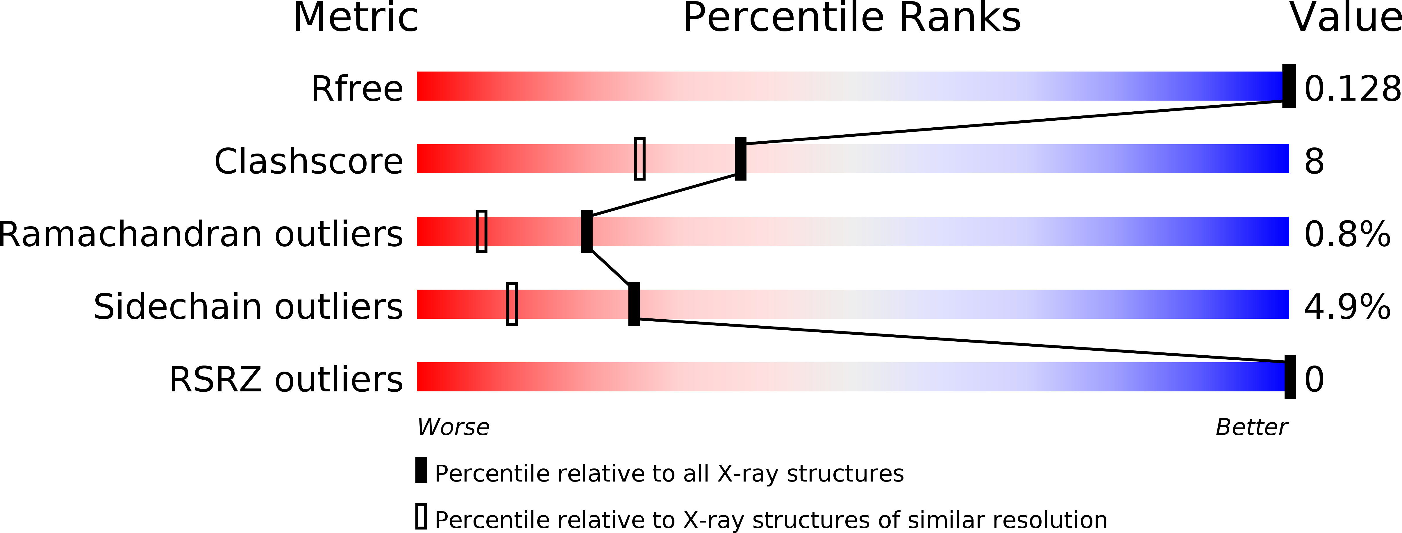
Resolution:
1.80 Å
R-Value Free:
0.14
R-Value Work:
0.12
R-Value Observed:
0.14
Space Group:
P 63
Macromolecular Entities
Structures with similar UniProt ID
Protein Blast
Polymer Type:polypeptide(L)
Molecule:Photoactive yellow protein
Gene (Uniprot):pyp
Chain IDs:A
Chain Length:125
Number of Molecules:1
Biological Source:Halorhodospira halophila
Ligand Molecules
Primary Citation
pH Dependence of the Photoactive Yellow Protein Photocycle Investigated by Time-Resolved Crystallography.
Biophys.J. 102 325 332 (2012)
PMID: 22339869 DOI: 10.1016/j.bpj.2011.11.4021

Abstact

Visualizing the three-dimensional structures of a protein during its biological activity is key to understanding its mechanism. In general, protein structure and function are pH-dependent. Changing the pH provides new insights into the mechanisms that are involved in protein activity. Photoactive yellow protein (PYP) is a signaling protein that serves as an ideal model for time-dependent studies on light-activated proteins. Its photocycle is studied extensively under different pH conditions. However, the structures of the intermediates remain unknown until time-resolved crystallography is employed. With the newest beamline developments, a comprehensive time series of Laue data can now be collected from a single protein crystal. This allows us to vary the pH. Here we present the first structure, to our knowledge, of a short-lived protein-inhibitor complex formed in the pB state of the PYP photocycle at pH 4. A water molecule that is transiently stabilized in the chromophore active site prevents the relaxation of the chromophore back to the trans configuration. As a result, the dark-state recovery is slowed down dramatically. At pH 9, PYP stops cycling through the pB state altogether. The electrostatic environment in the chromophore-binding site is the likely reason for this altered kinetics at different pH values.

Legend

Protein

Chemical

Disease

Primary Citation of related structures
Feedback Form
Name
Email
Institute
Feedback