3ULP image
Deposition Date 2011-11-11
Release Date 2012-05-09
Last Version Date 2023-09-13
Entry Detail
PDB ID:
3ULP
Title:
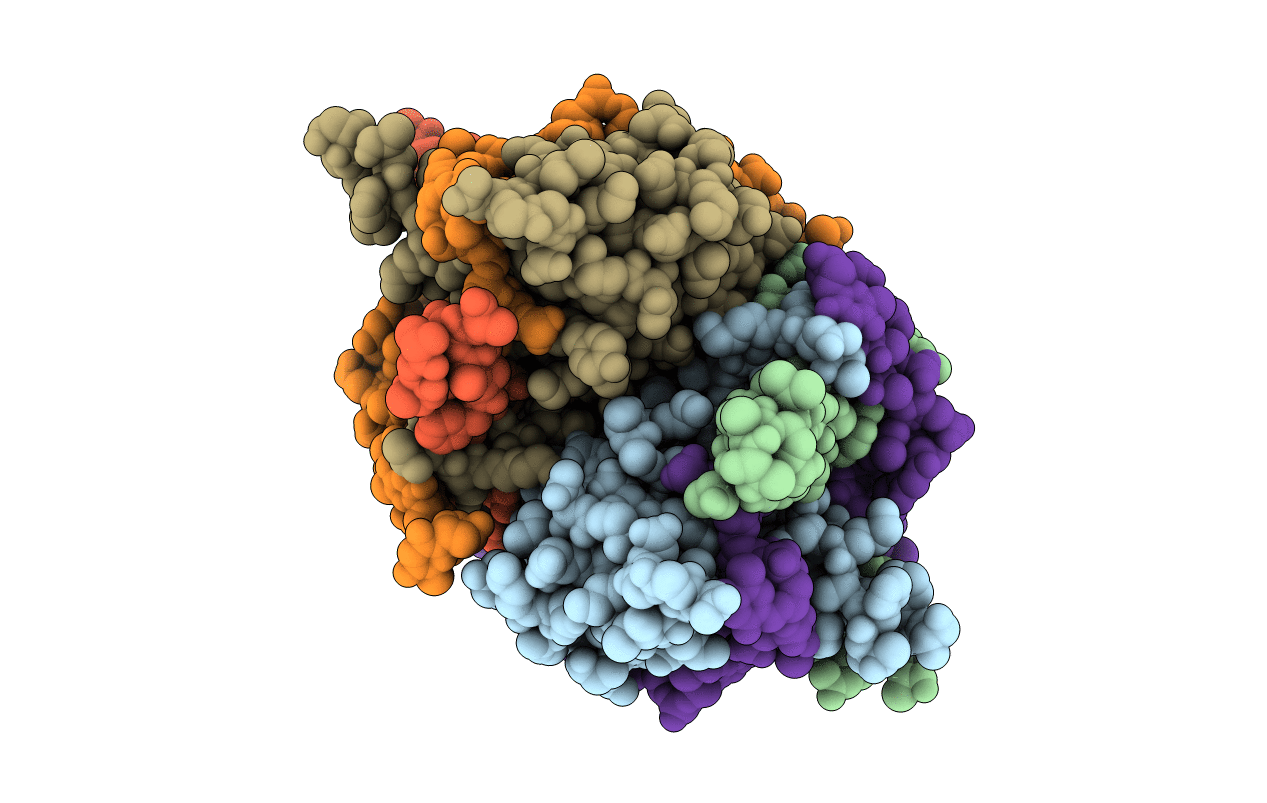
Plasmodium falciparum SSB complex with ssDNA
Biological Source:
Source Organism:
Host Organism:
Method Details:
Experimental Method:
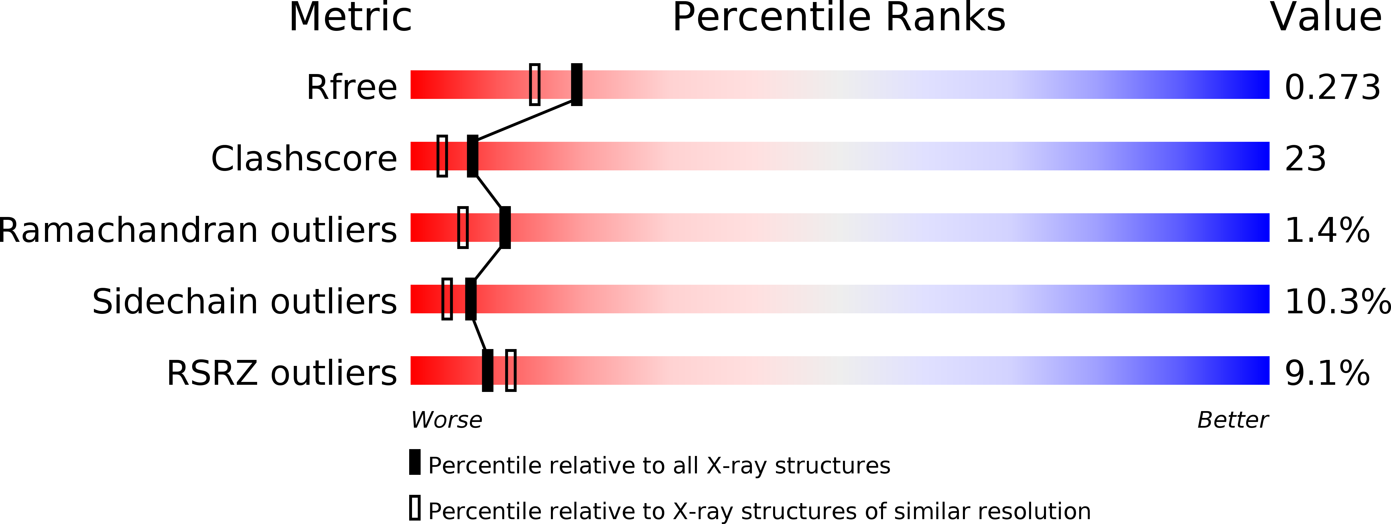
Resolution:
2.10 Å
R-Value Free:
0.27
R-Value Work:
0.21
R-Value Observed:
0.22
Space Group:
C 1 2 1
Macromolecular Entities
Structures with similar UniProt ID
Protein Blast
Polymer Type:polypeptide(L)
Molecule:Single-strand binding protein
Gene (Uniprot):PF3D7_0508800
Chain IDs:A, B, C, D
Chain Length:124
Number of Molecules:4
Biological Source:Plasmodium falciparum
Polymer Type:polydeoxyribonucleotide
Molecule:DNA (35-MER)
Chain IDs:E (auth: R), F (auth: Q)
Chain Length:35
Number of Molecules:2
Biological Source:Synthetic DNA
Primary Citation
Plasmodium falciparum SSB Tetramer Wraps Single-Stranded DNA with Similar Topology but Opposite Polarity to E. coli SSB.
J.Mol.Biol. 420 269 283 (2012)
PMID: 22543099 DOI: 10.1016/j.jmb.2012.04.021

Abstact

Single-stranded DNA binding (SSB) proteins play central roles in genome maintenance in all organisms. Plasmodium falciparum, the causative agent of malaria, encodes an SSB protein that localizes to the apicoplast and likely functions in the replication and maintenance of its genome. P. falciparum SSB (Pf-SSB) shares a high degree of sequence homology with bacterial SSB proteins but differs in the composition of its C-terminus, which interacts with more than a dozen other proteins in Escherichia coli SSB (Ec-SSB). Using sedimentation methods, we show that Pf-SSB forms a stable homo-tetramer alone and when bound to single-stranded DNA (ssDNA). We also present a crystal structure at 2.1 Å resolution of the Pf-SSB tetramer bound to two (dT)(35) molecules. The Pf-SSB tetramer is structurally similar to the Ec-SSB tetramer, and ssDNA wraps completely around the tetramer with a "baseball seam" topology that is similar to Ec-SSB in its "65 binding mode". However, the polarity of the ssDNA wrapping around Pf-SSB is opposite to that observed for Ec-SSB. The interactions between the bases in the DNA and the amino acid side chains also differ from those observed in the Ec-SSB-DNA structure, suggesting that other differences may exist in the DNA binding properties of these structurally similar proteins.

Legend

Protein

Chemical

Disease

Primary Citation of related structures
Feedback Form
Name
Email
Institute
Feedback