Deposition Date
1996-12-11
Release Date
1997-10-15
Last Version Date
2024-02-28
Entry Detail
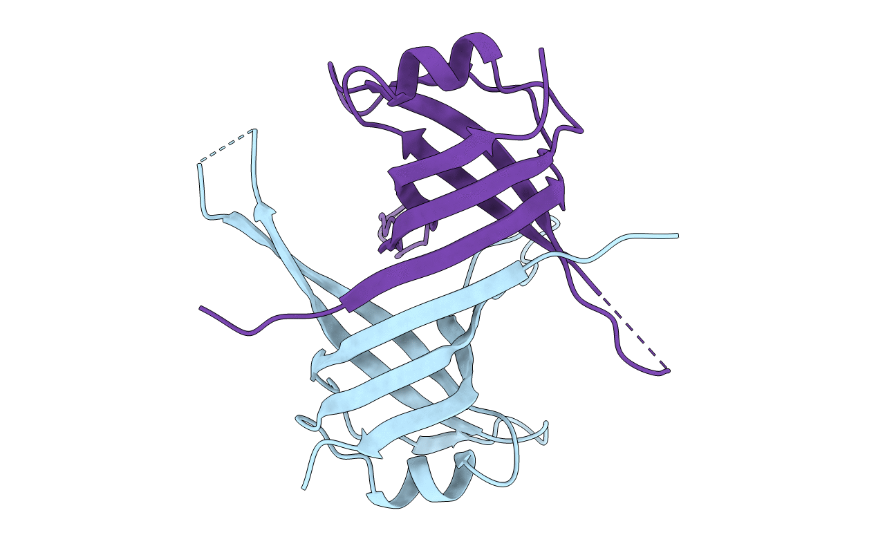
PDB ID:
3ULL
Keywords:
Title:
HUMAN MITOCHONDRIAL SINGLE-STRANDED DNA BINDING PROTEIN
Biological Source:
Source Organism(s):
Homo sapiens (Taxon ID: 9606)
Method Details:
Experimental Method:
Resolution:
2.40 Å
R-Value Free:
0.23
R-Value Work:
0.19
R-Value Observed:
0.19
Space Group:
P 41 21 2