3ULF image
Deposition Date 2011-11-10
Release Date 2012-04-11
Last Version Date 2024-11-06
Entry Detail
PDB ID:
3ULF
Title:
The light state structure of the blue-light photoreceptor Aureochrome1 LOV
Biological Source:
Source Organism:
Host Organism:
Method Details:
Experimental Method:
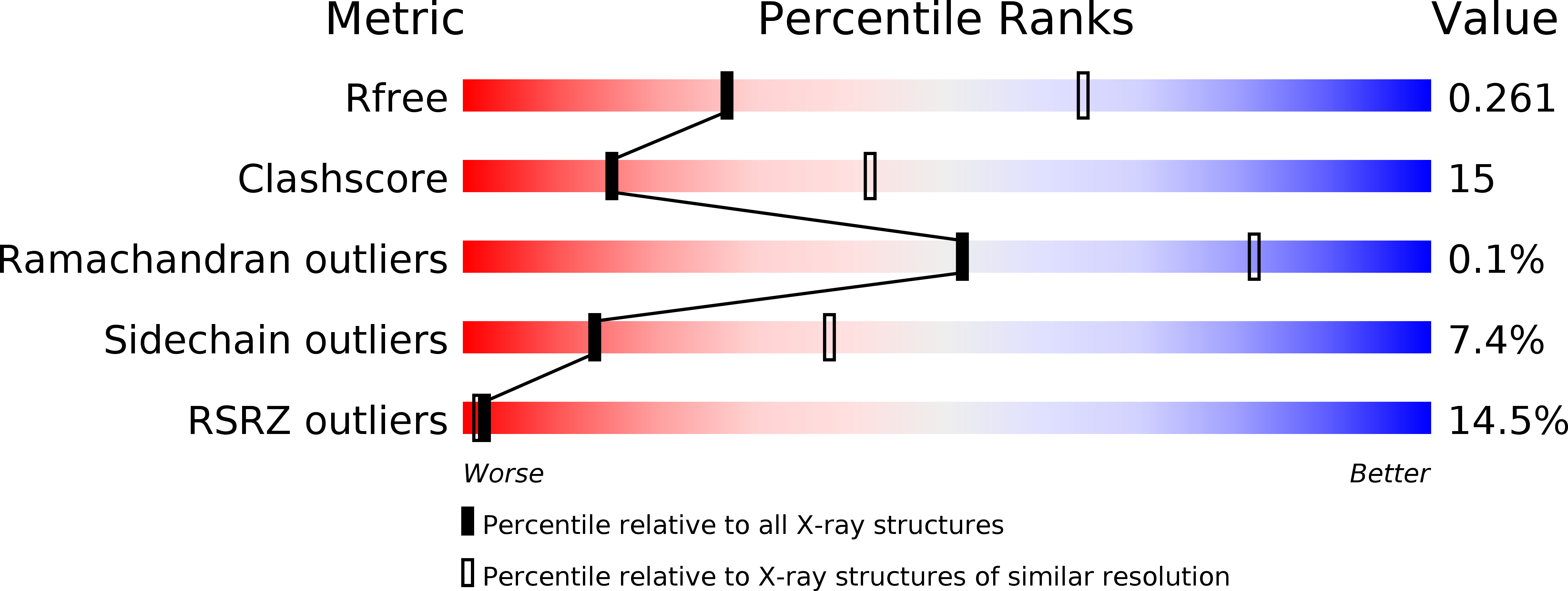
Resolution:
2.90 Å
R-Value Free:
0.26
R-Value Work:
0.20
R-Value Observed:
0.20
Space Group:
P 43
Macromolecular Entities
Structures with similar UniProt ID
Protein Blast
Polymer Type:polypeptide(L)
Molecule:Aureochrome1
Gene (Uniprot):AUREO1
Chain IDs:A, B, C, D, E, F
Chain Length:170
Number of Molecules:6
Biological Source:Vaucheria frigida
Primary Citation
Crystal structures of Aureochrome1 LOV suggest new design strategies for optogenetics.
Structure 20 698 706 (2012)
PMID: 22483116 DOI: 10.1016/j.str.2012.02.016

Abstact

Aureochrome1, a signaling photoreceptor from a eukaryotic photosynthetic stramenopile, confers blue-light-regulated DNA binding on the organism. Its topology, in which a C-terminal LOV sensor domain is linked to an N-terminal DNA-binding bZIP effector domain, contrasts with the reverse sensor-effector topology in most other known LOV-photoreceptors. How, then, is signal transmitted in Aureochrome1? The dark- and light-state crystal structures of Aureochrome1 LOV domain (AuLOV) show that its helical N- and C-terminal flanking regions are packed against the external surface of the core β sheet, opposite to the FMN chromophore on the internal surface. Light-induced conformational changes occur in the quaternary structure of the AuLOV dimer and in Phe298 of the Hβ strand in the core. The properties of AuLOV extend the applicability of LOV domains as versatile design modules that permit fusion to effector domains via either the N- or C-termini to confer blue-light sensitivity.

Legend

Protein

Chemical

Disease

Primary Citation of related structures