Deposition Date
2011-11-10
Release Date
2012-11-28
Last Version Date
2023-09-13
Entry Detail
PDB ID:
3UL5
Keywords:
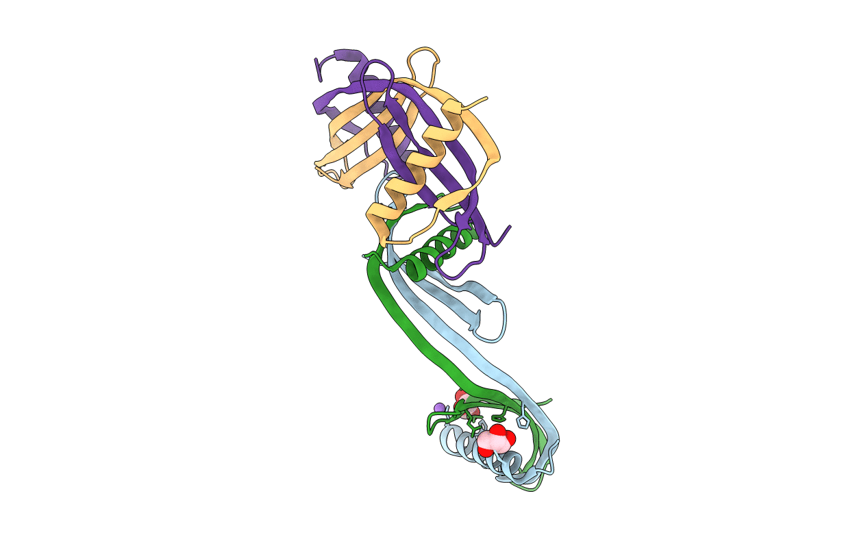
Title:
Saccharum officinarum canecystatin-1 in space group C2221
Biological Source:
Source Organism(s):
Saccharum officinarum (Taxon ID: 4547)
Expression System(s):
Method Details:
Experimental Method:
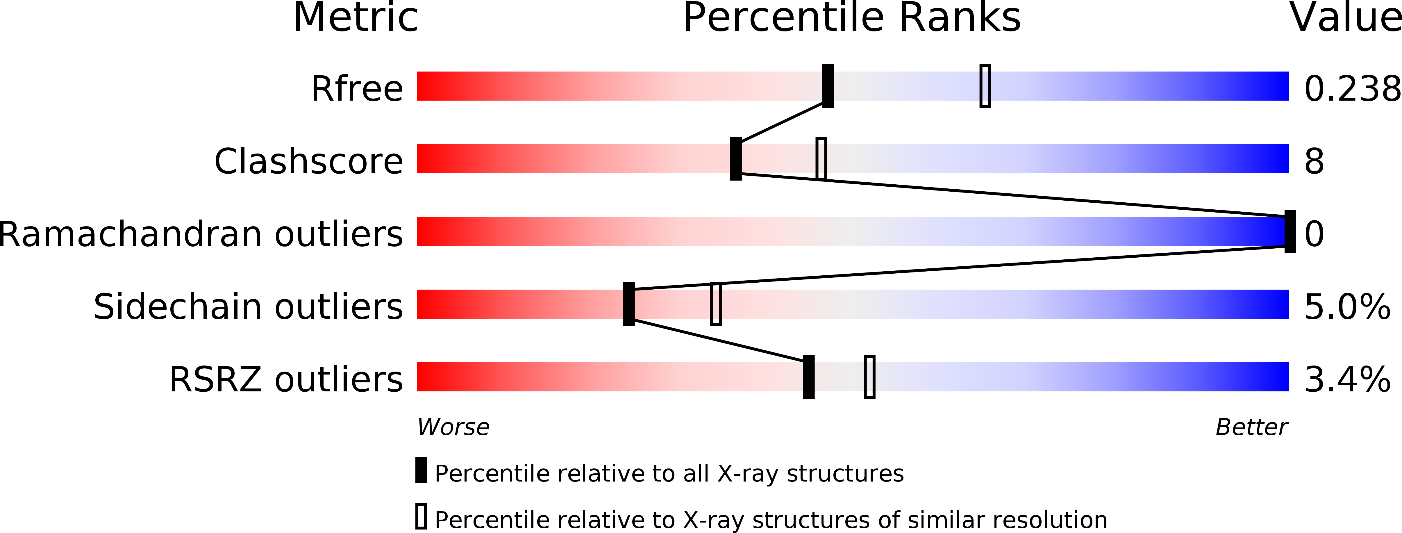
Resolution:
2.30 Å
R-Value Free:
0.25
R-Value Work:
0.22
R-Value Observed:
0.22
Space Group:
C 2 2 21