Deposition Date
2011-11-08
Release Date
2012-07-25
Last Version Date
2024-11-20
Entry Detail
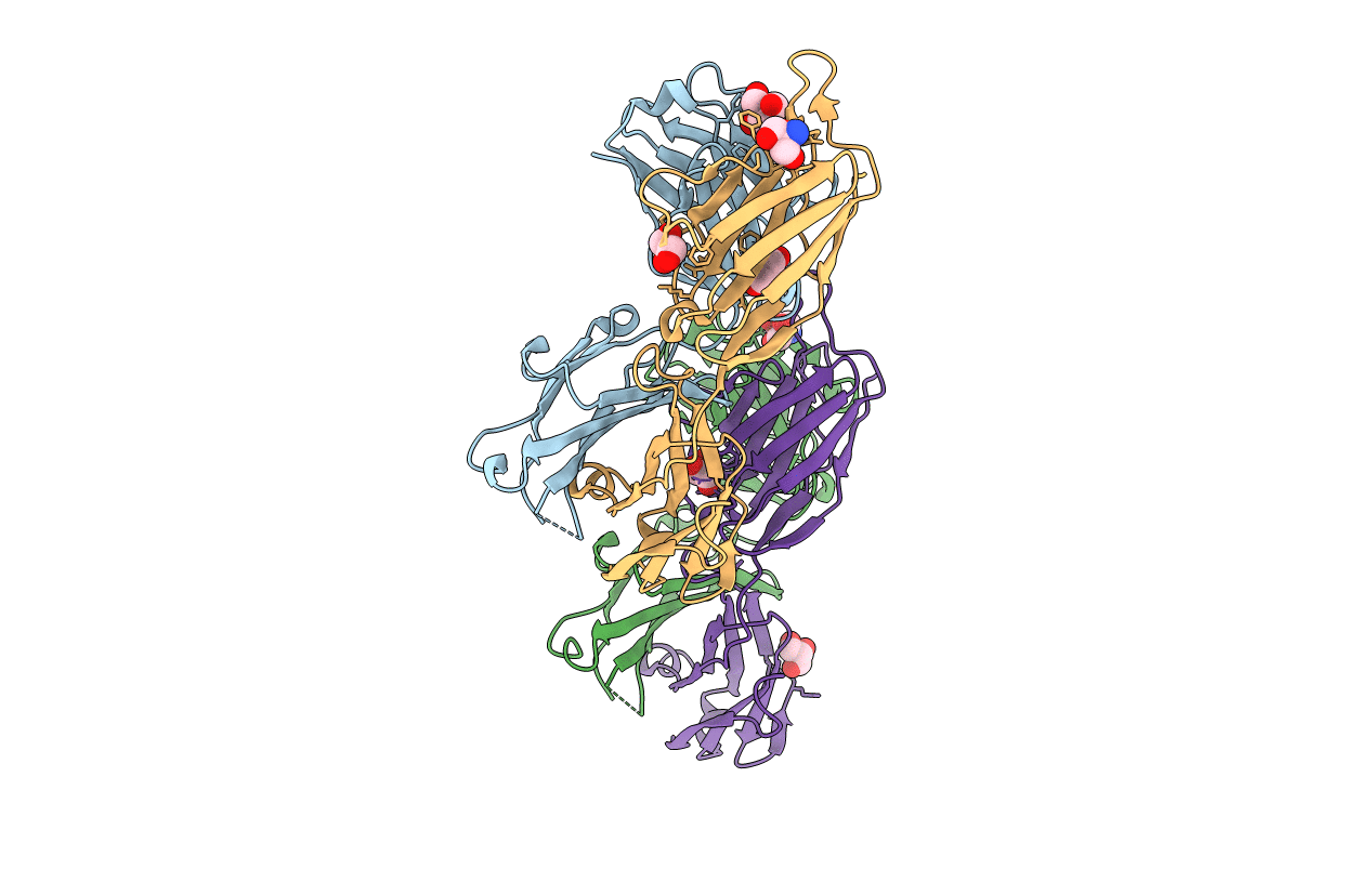
PDB ID:
3UJT
Keywords:
Title:
Structure of the Fab fragment of Ab-52, an antibody that binds the O-antigen of Francisella tularensis
Biological Source:
Source Organism(s):
Mus musculus (Taxon ID: 10090)
Method Details:
Experimental Method:
Resolution:
2.10 Å
R-Value Free:
0.21
R-Value Work:
0.18
R-Value Observed:
0.18
Space Group:
P 1 21 1