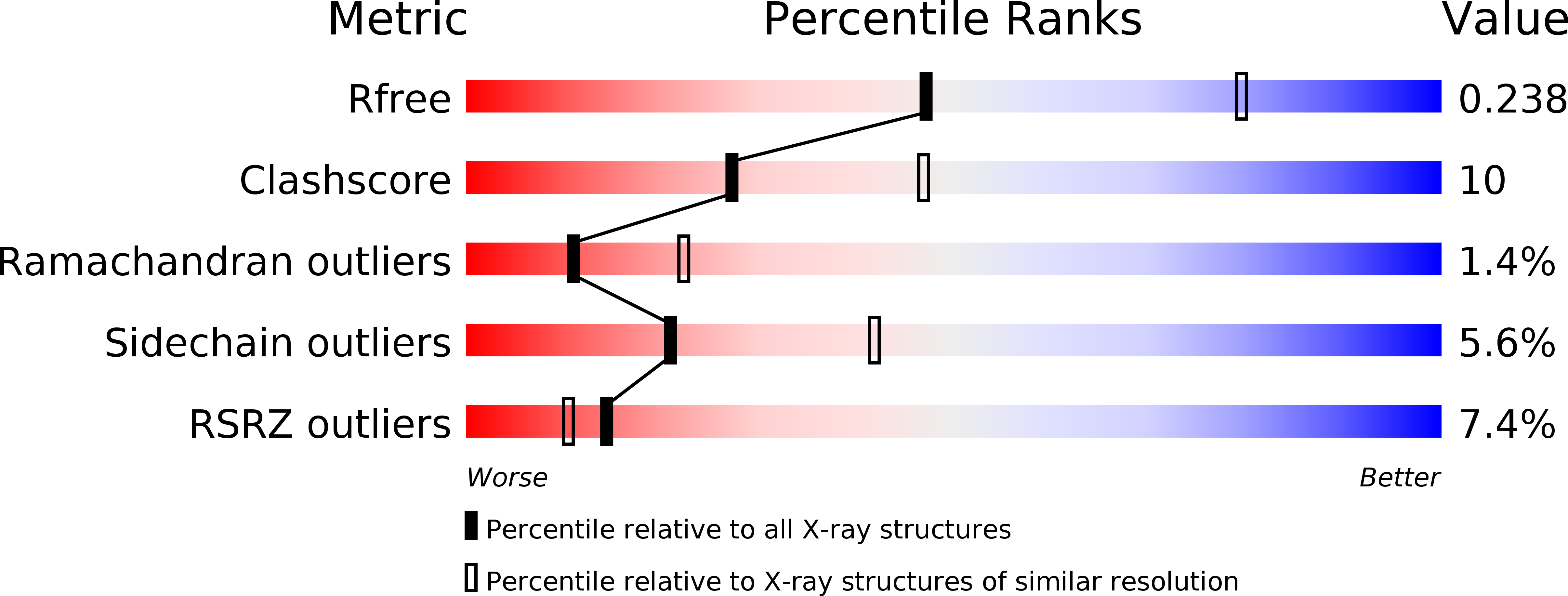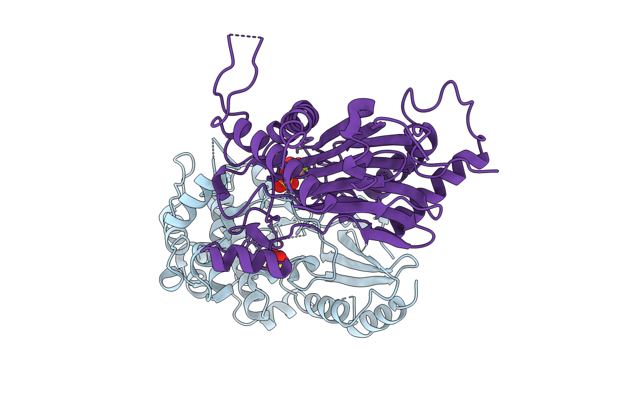
3UJG image
Deposition Date 2011-11-07
Release Date 2012-02-15
Last Version Date 2024-02-28
Entry Detail
PDB ID:
3UJG
Title:
Crystal structure of SnRK2.6 in complex with HAB1
Biological Source:
Source Organism:
Host Organism:
Method Details:
Experimental Method:
Resolution:
2.60 Å
R-Value Free:
0.23
R-Value Work:
0.20
R-Value Observed:
0.20
Space Group:
I 4
Macromolecular Entities
Polymer Type:polypeptide(L)
Molecule:Serine/threonine-protein kinase SRK2E
Gene (Uniprot):SRK2E
Mutations:D296A, E297A
Chain IDs:A
Chain Length:361
Number of Molecules:1
Biological Source:Arabidopsis thaliana
Polymer Type:polypeptide(L)
Molecule:Protein phosphatase 2C 16
Gene (Uniprot):HAB1
Chain IDs:B
Chain Length:350
Number of Molecules:1
Biological Source:Arabidopsis thaliana
Primary Citation
Molecular mimicry regulates ABA signaling by SnRK2 kinases and PP2C phosphatases.
Science 335 85 88 (2012)
PMID: 22116026 DOI: 10.1126/science.1215106

Abstact

Abscisic acid (ABA) is an essential hormone for plants to survive environmental stresses. At the center of the ABA signaling network is a subfamily of type 2C protein phosphatases (PP2Cs), which form exclusive interactions with ABA receptors and subfamily 2 Snfl-related kinase (SnRK2s). Here, we report a SnRK2-PP2C complex structure, which reveals marked similarity in PP2C recognition by SnRK2 and ABA receptors. In the complex, the kinase activation loop docks into the active site of PP2C, while the conserved ABA-sensing tryptophan of PP2C inserts into the kinase catalytic cleft, thus mimicking receptor-PP2C interactions. These structural results provide a simple mechanism that directly couples ABA binding to SnRK2 kinase activation and highlight a new paradigm of kinase-phosphatase regulation through mutual packing of their catalytic sites.

Legend

Protein

Chemical

Disease

Primary Citation of related structures