3UHR image
Entry Detail
PDB ID:
3UHR
Title:
HBI (L36F) deoxy
Biological Source:
Source Organism:
Host Organism:
PDB Version:
Deposition Date:
2011-11-03
Release Date:
2011-12-28
Method Details:
Experimental Method:
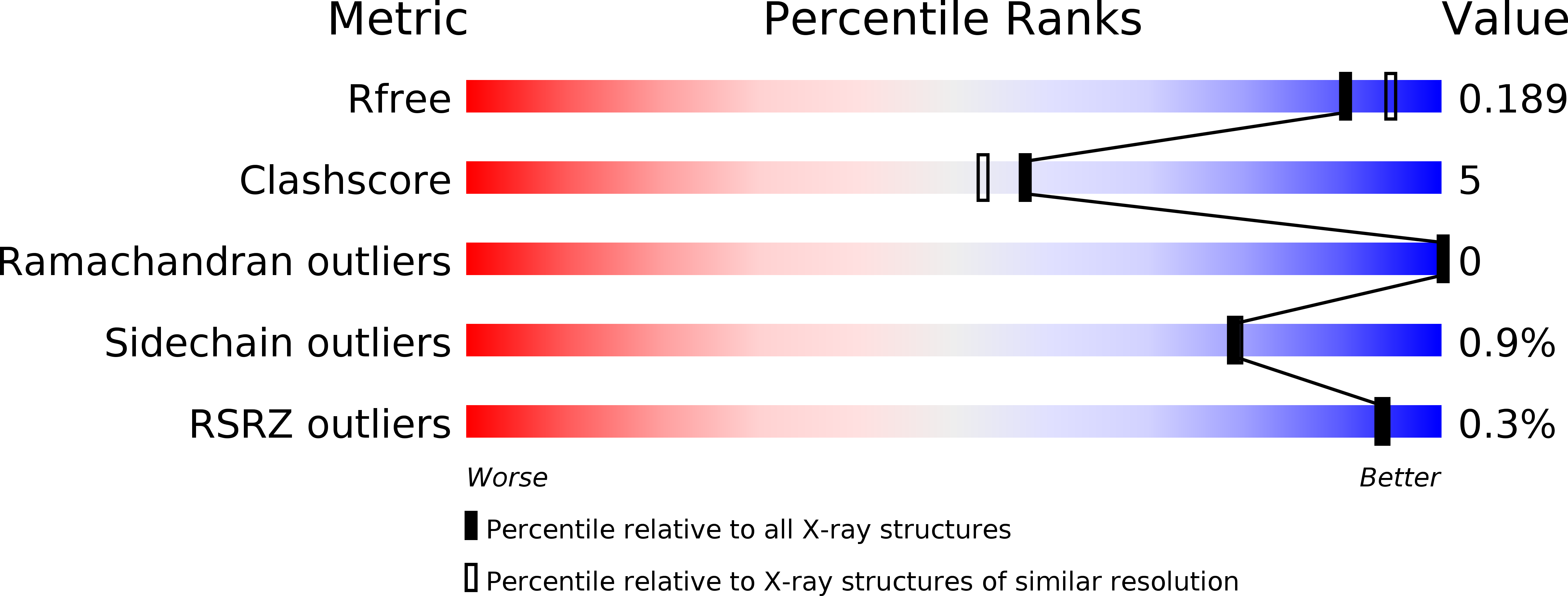
Resolution:
1.90 Å
R-Value Free:
0.19
R-Value Work:
0.16
R-Value Observed:
0.16
Space Group:
C 2 2 21
Macromolecular Entities
Polymer Type:polypeptide(L)
Description:Globin-1
Mutations:L36F
Chain IDs:A, B
Chain Length:146
Number of Molecules:2
Biological Source:Scapharca inaequivalvis
Ligand Molecules
Primary Citation
Cooperative macromolecular device revealed by meta-analysis of static and time-resolved structures.
Proc.Natl.Acad.Sci.USA 109 107 112 (2012)
PMID: 22171006 DOI: 10.1073/pnas.1109213108

Abstact

Here we present a meta-analysis of a large collection of static structures of a protein in the Protein Data Bank in order to extract the progression of structural events during protein function. We apply this strategy to the homodimeric hemoglobin HbI from Scapharca inaequivalvis. We derive a simple dynamic model describing how binding of the first ligand in one of the two chemically identical subunits facilitates a second binding event in the other partner subunit. The results of our ultrafast time-resolved crystallographic studies support this model. We demonstrate that HbI functions like a homodimeric mechanical device, such as pliers or scissors. Ligand-induced motion originating in one subunit is transmitted to the other via conserved pivot points, where the E and F' helices from two partner subunits are "bolted" together to form a stable dimer interface permitting slight relative rotation but preventing sliding.

Legend

Protein

Chemical

Disease