3UGM image
Deposition Date 2011-11-02
Release Date 2012-01-04
Last Version Date 2024-02-28
Entry Detail
PDB ID:
3UGM
Title:
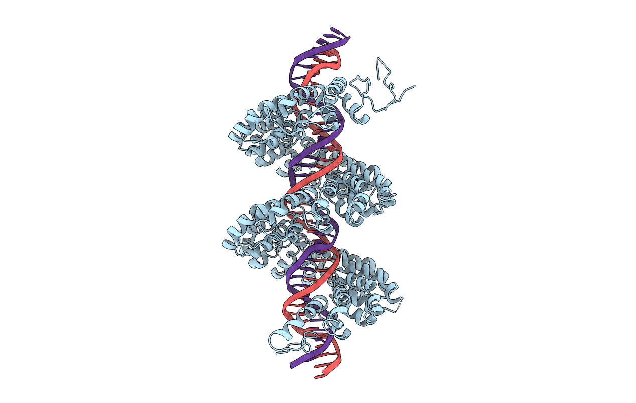
Structure of TAL effector PthXo1 bound to its DNA target
Biological Source:
Source Organism:
Xanthomonas oryzae (Taxon ID: 360094)
(Taxon ID: )
Host Organism:
Method Details:
Experimental Method:
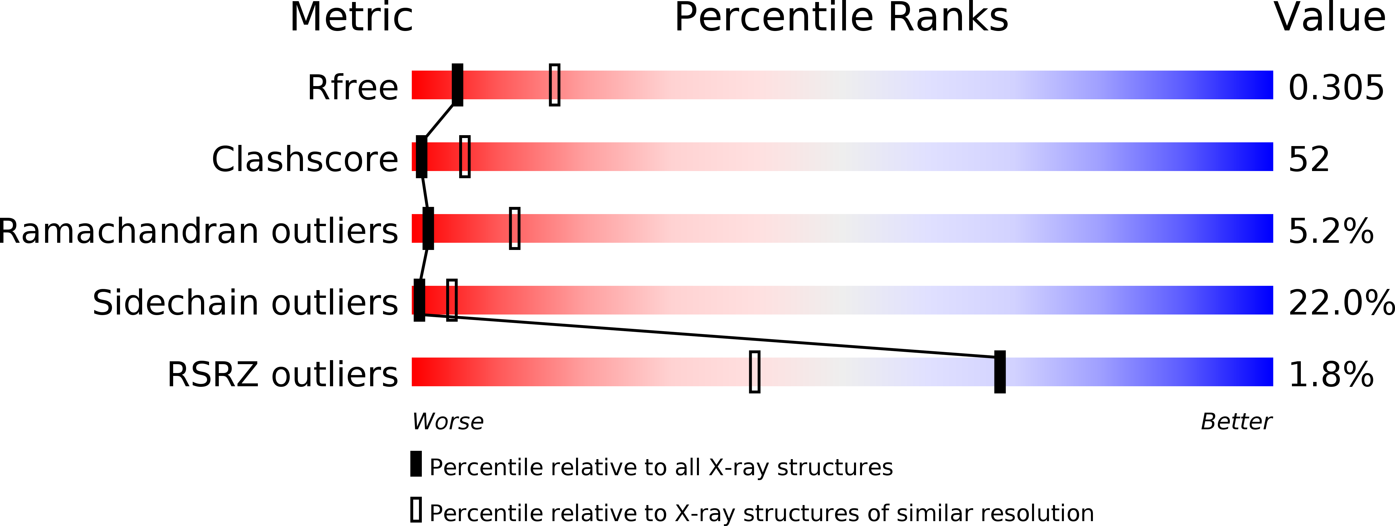
Resolution:
3.00 Å
R-Value Free:
0.29
R-Value Work:
0.26
Space Group:
P 21 21 21
Macromolecular Entities
Polymer Type:polypeptide(L)
Molecule:TAL effector AvrBs3/PthA
Gene (Uniprot):pthXo1
Chain IDs:A
Chain Length:1047
Number of Molecules:1
Biological Source:Xanthomonas oryzae
Polymer Type:polydeoxyribonucleotide
Molecule:DNA-1
Chain IDs:B
Chain Length:38
Number of Molecules:1
Biological Source:
Polymer Type:polydeoxyribonucleotide
Molecule:DNA-2
Chain IDs:C
Chain Length:38
Number of Molecules:1
Biological Source:
Primary Citation
The Crystal Structure of TAL Effector PthXo1 Bound to Its DNA Target.
Science 335 716 719 (2012)
PMID: 22223736 DOI: 10.1126/science.1216211

Abstact

DNA recognition by TAL effectors is mediated by tandem repeats, each 33 to 35 residues in length, that specify nucleotides via unique repeat-variable diresidues (RVDs). The crystal structure of PthXo1 bound to its DNA target was determined by high-throughput computational structure prediction and validated by heavy-atom derivatization. Each repeat forms a left-handed, two-helix bundle that presents an RVD-containing loop to the DNA. The repeats self-associate to form a right-handed superhelix wrapped around the DNA major groove. The first RVD residue forms a stabilizing contact with the protein backbone, while the second makes a base-specific contact to the DNA sense strand. Two degenerate amino-terminal repeats also interact with the DNA. Containing several RVDs and noncanonical associations, the structure illustrates the basis of TAL effector-DNA recognition.

Legend

Protein

Chemical

Disease

Primary Citation of related structures