Deposition Date
2011-11-01
Release Date
2012-08-08
Last Version Date
2024-11-20
Entry Detail
PDB ID:
3UFK
Keywords:
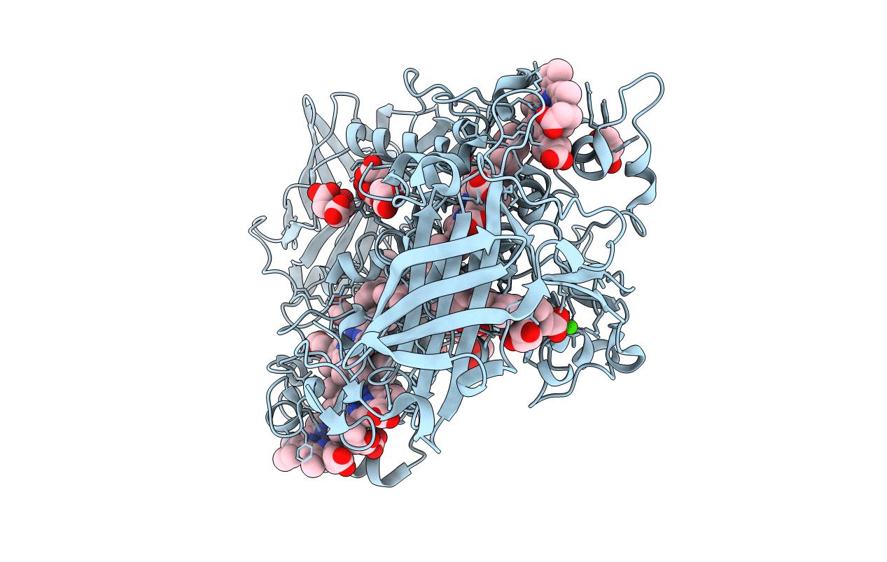
Title:
Crystal structure of UndA complexed with Iron Nitrilotriacetate
Biological Source:
Source Organism(s):
Shewanella sp. HRCR_06 (Taxon ID: 1043181)
Expression System(s):
Method Details:
Experimental Method:
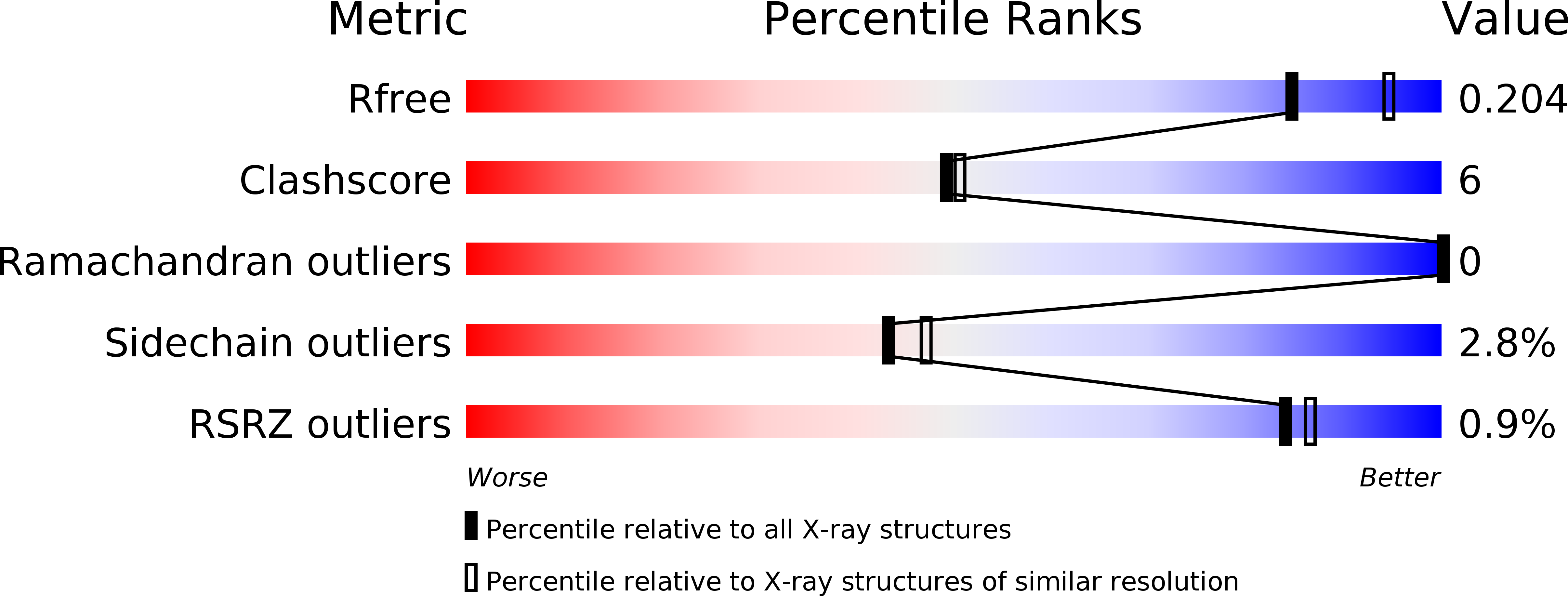
Resolution:
2.10 Å
R-Value Free:
0.20
R-Value Work:
0.15
R-Value Observed:
0.15
Space Group:
P 21 21 21