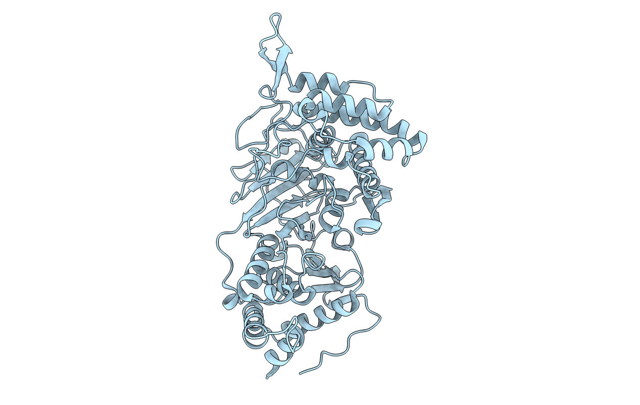
3UEK image
Entry Detail
PDB ID:
3UEK
Keywords:
Title:
Crystal structure of the catalytic domain of rat poly (ADP-ribose) glycohydrolase
Biological Source:
Source Organism:
Host Organism:
PDB Version:
Deposition Date:
2011-10-30
Release Date:
2012-05-23
Method Details:
Experimental Method:
Resolution:
1.95 Å
R-Value Free:
0.21
R-Value Work:
0.18
R-Value Observed:
0.18
Space Group:
C 2 2 21
Macromolecular Entities
Polymer Type:polypeptide(L)
Description:Poly(ADP-ribose) glycohydrolase
Chain IDs:A
Chain Length:588
Number of Molecules:1
Biological Source:Rattus norvegicus
Primary Citation
Structure of mammalian poly(ADP-ribose) glycohydrolase reveals a flexible tyrosine clasp as a substrate-binding element.
Nat.Struct.Mol.Biol. 19 653 656 (2012)
PMID: 22609859 DOI: 10.1038/nsmb.2305

Abstact

Reversible post-translational modification by poly(ADP-ribose) (PAR) regulates chromatin structure, DNA repair and cell fate in response to genotoxic stress. PAR glycohydrolase (PARG) removes PAR chains from poly ADP-ribosylated proteins to restore protein function and release oligo(ADP-ribose) chains to signal damage. Here we report crystal structures of mammalian PARG and its complex with a substrate mimic that reveal an open substrate-binding site and a unique 'tyrosine clasp' enabling endoglycosidic cleavage of branched PAR chains.

Legend

Protein

Chemical

Disease

Primary Citation of related structures