3UDG image
Deposition Date 2011-10-28
Release Date 2012-05-16
Last Version Date 2024-02-28
Entry Detail
PDB ID:
3UDG
Title:
Structure of Deinococcus radiodurans SSB bound to ssDNA
Biological Source:
Source Organism:
Host Organism:
Method Details:
Experimental Method:
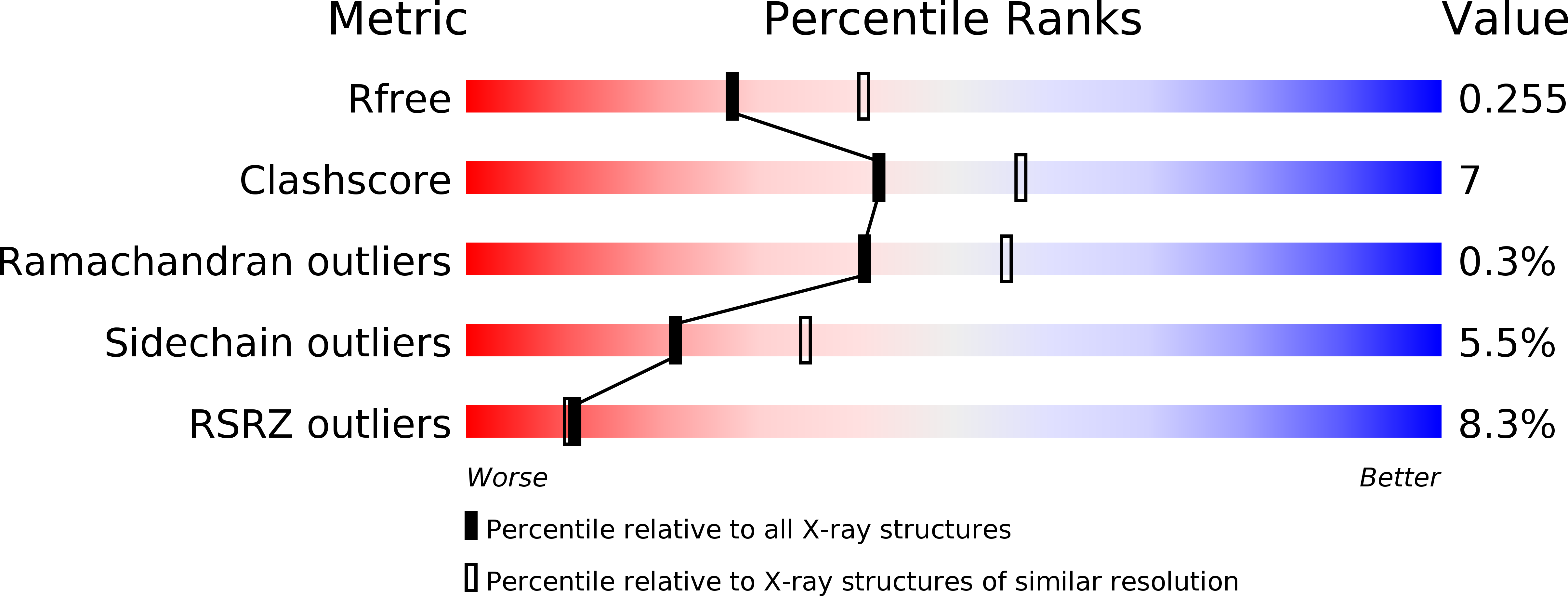
Resolution:
2.40 Å
R-Value Free:
0.26
R-Value Work:
0.20
R-Value Observed:
0.20
Space Group:
C 1 2 1
Macromolecular Entities
Polymer Type:polypeptide(L)
Molecule:Single-stranded DNA-binding protein
Gene (Uniprot):ssb
Chain IDs:A, B, C
Chain Length:301
Number of Molecules:3
Biological Source:Deinococcus radiodurans
Polymer Type:polydeoxyribonucleotide
Molecule:5'-D(P*TP*TP*TP*TP*TP*TP*TP*TP*TP*TP*TP*TP*TP*TP*TP*TP*TP*TP*TP*TP*TP*TP*TP*TP*TP*TP*TP*TP*TP*TP*TP*TP*TP*TP*T)-3'
Chain IDs:D, E, F, G, H, I
Chain Length:35
Number of Molecules:6
Biological Source:Synthetic DNA
Ligand Molecules
Primary Citation
Structure and Cellular Dynamics of Deinococcus radiodurans Single-stranded DNA (ssDNA)-binding Protein (SSB)-DNA Complexes.
J.Biol.Chem. 287 22123 22132 (2012)
PMID: 22570477 DOI: 10.1074/jbc.M112.367573

Abstact

The single-stranded DNA (ssDNA)-binding protein from the radiation-resistant bacterium Deinococcus radiodurans (DrSSB) functions as a homodimer in which each monomer contains two oligonucleotide-binding (OB) domains. This arrangement is exceedingly rare among bacterial SSBs, which typically form homotetramers of single-OB domain subunits. To better understand how this unusual structure influences the DNA binding and biological functions of DrSSB in D. radiodurans radiation resistance, we have examined the structure of DrSSB in complex with ssDNA and the DNA damage-dependent cellular dynamics of DrSSB. The x-ray crystal structure of the DrSSB-ssDNA complex shows that ssDNA binds to surfaces of DrSSB that are analogous to those mapped in homotetrameric SSBs, although there are distinct contacts in DrSSB that mediate species-specific ssDNA binding. Observations by electron microscopy reveal two salt-dependent ssDNA-binding modes for DrSSB that strongly resemble those of the homotetrameric Escherichia coli SSB, further supporting a shared overall DNA binding mechanism between the two classes of bacterial SSBs. In vivo, DrSSB levels are heavily induced following exposure to ionizing radiation. This accumulation is accompanied by dramatic time-dependent DrSSB cellular dynamics in which a single nucleoid-centric focus of DrSSB is observed within 1 h of irradiation but is dispersed by 3 h after irradiation. These kinetics parallel those of D. radiodurans postirradiation genome reconstitution, suggesting that DrSSB dynamics could play important organizational roles in DNA repair.

Legend

Protein

Chemical

Disease

Primary Citation of related structures