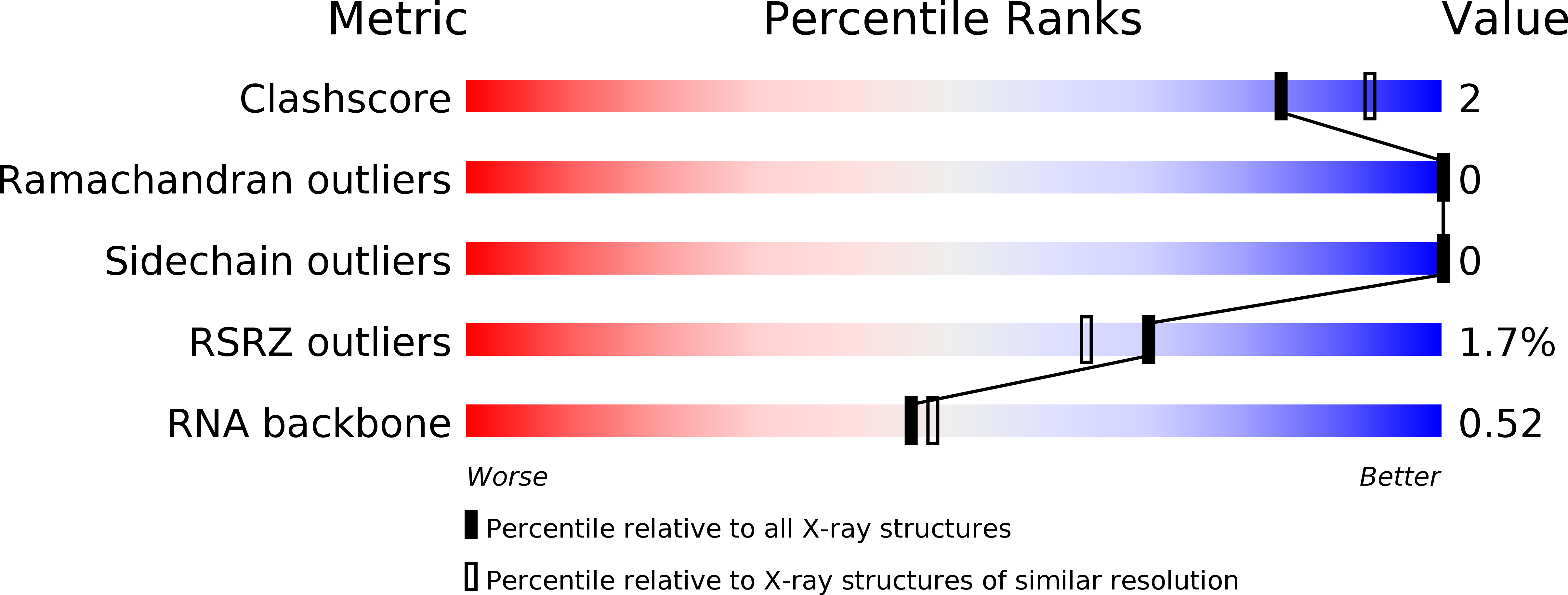
3UCU image
Deposition Date 2011-10-27
Release Date 2012-01-04
Last Version Date 2024-02-28
Entry Detail
PDB ID:
3UCU
Title:
The c-di-GMP-I riboswitch bound to pGpG
Biological Source:
Source Organism:
Homo sapiens (Taxon ID: 9606)
Host Organism:
Method Details:
Experimental Method:
Resolution:
2.80 Å
R-Value Free:
0.26
R-Value Work:
0.21
R-Value Observed:
0.21
Space Group:
P 1 21 1
Macromolecular Entities
Structures with similar UniProt ID
Protein Blast
Polymer Type:polypeptide(L)
Molecule:U1 small nuclear ribonucleoprotein A
Gene (Uniprot):SNRPA
Mutagens:Y31H, Q36R
Chain IDs:A (auth: P)
Chain Length:98
Number of Molecules:1
Biological Source:Homo sapiens
Polymer Type:polyribonucleotide
Molecule:RNA (92-MER)
Chain IDs:B (auth: R)
Chain Length:92
Number of Molecules:1
Biological Source:
Modified Residue
Compound ID Chain ID Parent Comp ID Details 2D Image
GTP B G GUANOSINE-5'-TRIPHOSPHATE
Ligand Molecules
Primary Citation
Structural and biochemical characterization of linear dinucleotide analogues bound to the c-di-GMP-I aptamer.
Biochemistry 51 425 432 (2012)
PMID: 22148472 DOI: 10.1021/bi2016662

Abstact

The cyclic dinucleotide c-di-GMP regulates lifestyle transitions in many bacteria, such as the change from a free motile state to a biofilm-forming community. Riboswitches that bind this second messenger are important downstream targets in this bacterial signaling pathway. The breakdown of c-di-GMP in the cell is accomplished enzymatically and results in the linear dinucleotide pGpG. The c-di-GMP-binding riboswitches must be able to discriminate between their cognate cyclic ligand and linear dinucleotides in order to be selective biological switches. It has been reported that the c-di-GMP-I riboswitch binds c-di-GMP 5 orders of magnitude better than the linear pGpG, but the cause of this large energetic difference in binding is unknown. Here we report binding data and crystal structures of several linear c-di-GMP analogues in complex with the c-di-GMP-I riboswitch. These data reveal the parameters for phosphate recognition and the structural basis of linear dinucleotide binding to the riboswitch. Additionally, the pH dependence of binding shows that exclusion of pGpG is not due to the additional negative charge on the ligand. These data reveal principles that, along with published work, will contribute to the design of c-di-GMP analogues with properties desirable for use as chemical tools and potential therapeutics.

Legend

Protein

Chemical

Disease

Primary Citation of related structures