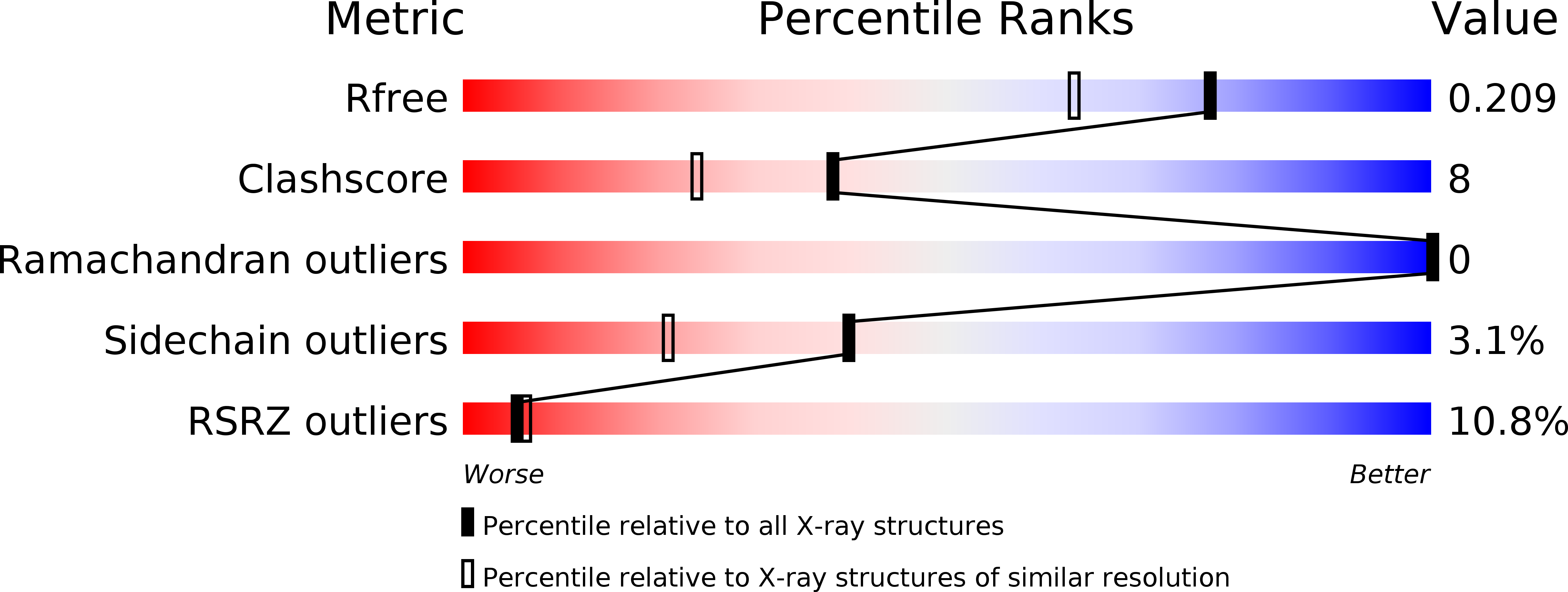
3UC5 image
Deposition Date 2011-10-26
Release Date 2012-11-07
Last Version Date 2024-11-27
Entry Detail
PDB ID:
3UC5
Keywords:
Title:
Phosphopantetheine adenylyltransferase from Mycobacterium tuberculosis complexed with ATP
Biological Source:
Source Organism:
Host Organism:
Method Details:
Experimental Method:
Resolution:
1.70 Å
R-Value Free:
0.22
R-Value Work:
0.17
R-Value Observed:
0.17
Space Group:
H 3 2
Macromolecular Entities
Polymer Type:polypeptide(L)
Molecule:Phosphopantetheine adenylyltransferase
Chain IDs:A
Chain Length:157
Number of Molecules:1
Biological Source:Mycobacterium tuberculosis
Ligand Molecules
Primary Citation
X-ray study of the conformational changes in the molecule of phosphopantetheine adenylyltransferase from Mycobacterium tuberculosis during the catalyzed reaction.
Acta Crystallogr.,Sect.D 68 1660 1670 (2012)
PMID: 23151631 DOI: 10.1107/S0907444912040206

Abstact

Structures of recombinant phosphopantetheine adenylyltransferase (PPAT) from Mycobacterium tuberculosis (PPATMt) in the apo form and in complex with the substrate ATP were determined at 1.62 and 1.70 Å resolution, respectively, using crystals grown in microgravity by the counter-diffusion method. The ATP molecule of the PPATMt-ATP complex was located with full occupancy in the active-site cavity. Comparison of the solved structures with previously determined structures of PPATMt complexed with the reaction product dephosphocoenzyme A (dPCoA) and the feedback inhibitor coenzyme A (CoA) was performed using superposition on C(α) atoms. The peculiarities of the arrangement of the ligands in the active-site cavity of PPATMt are described. The conformational states of the PPAT molecule in the consequent steps of the catalyzed reaction in the apo enzyme and the enzyme-substrate and enzyme-product complexes are characterized. It is shown that the binding of ATP and dPCoA induces the rearrangement of a short part of the polypeptide chain restricting the active-site cavity in the subunits of the hexameric enzyme molecule. The changes in the quaternary structure caused by this rearrangement are accompanied by a variation of the size of the inner water-filled channel which crosses the PPAT molecule along the threefold axis of the hexamer. The molecular mechanism of the observed changes is described.

Legend

Protein

Chemical

Disease

Primary Citation of related structures