Deposition Date
2011-10-25
Release Date
2011-11-16
Last Version Date
2024-11-20
Entry Detail
PDB ID:
3UBX
Keywords:
Title:
Crystal structure of the mouse CD1d-C20:2-aGalCer-L363 mAb Fab complex
Biological Source:
Source Organism(s):
Mus musculus (Taxon ID: 10090)
Expression System(s):
Method Details:
Experimental Method:
Resolution:
3.10 Å
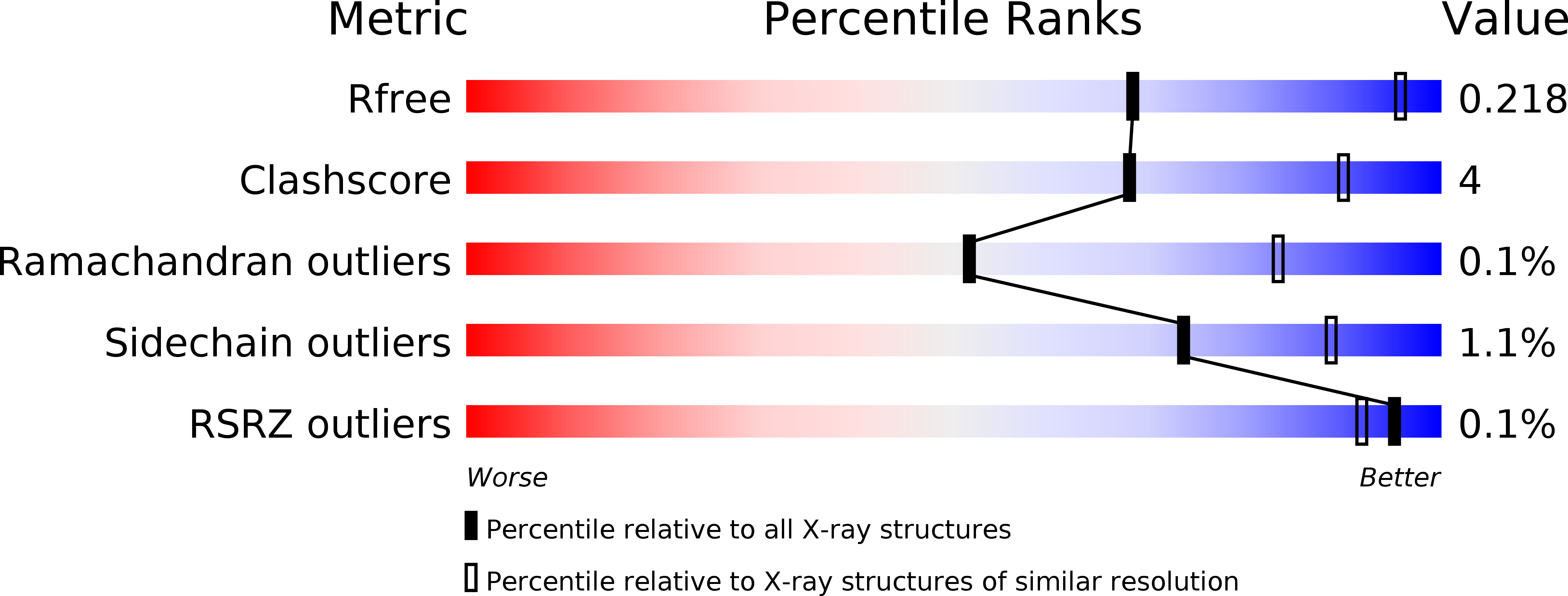
R-Value Free:
0.23
R-Value Work:
0.20
R-Value Observed:
0.20
Space Group:
P 63