
Deposition Date
2011-10-24
Release Date
2012-04-25
Last Version Date
2024-10-30
Entry Detail
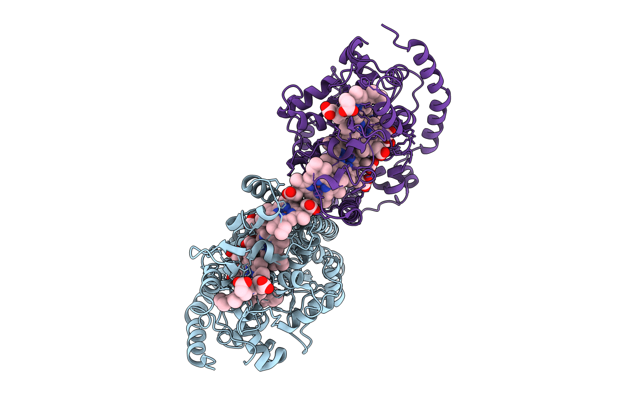
PDB ID:
3UBR
Keywords:
Title:
Laue structure of Shewanella oneidensis cytochrome-c Nitrite Reductase
Biological Source:
Source Organism(s):
Shewanella oneidensis (Taxon ID: 70863)
Expression System(s):
Method Details:
Experimental Method:
Resolution:
2.59 Å
R-Value Free:
0.28
R-Value Work:
0.19
R-Value Observed:
0.19
Space Group:
P 21 21 21
