
Deposition Date
2011-10-23
Release Date
2012-06-27
Last Version Date
2024-02-28
Entry Detail
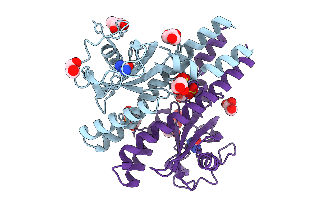
PDB ID:
3UB6
Keywords:
Title:
Periplasmic portion of the Helicobacter pylori chemoreceptor TlpB with urea bound
Biological Source:
Source Organism(s):
Helicobacter pylori (Taxon ID: 102617)
Expression System(s):
Method Details:
Experimental Method:
Resolution:
1.38 Å
R-Value Free:
0.14
R-Value Work:
0.12
R-Value Observed:
0.12
Space Group:
P 21 21 21
