Deposition Date
2011-10-18
Release Date
2012-10-31
Last Version Date
2024-10-09
Entry Detail
PDB ID:
3U99
Keywords:
Title:
The experimental X-ray structure of the new diheme cytochrome type c from Shewanella baltica OS155 sb-DHC
Biological Source:
Source Organism(s):
Shewanella baltica (Taxon ID: 325240)
Expression System(s):
Method Details:
Experimental Method:
Resolution:
1.15 Å
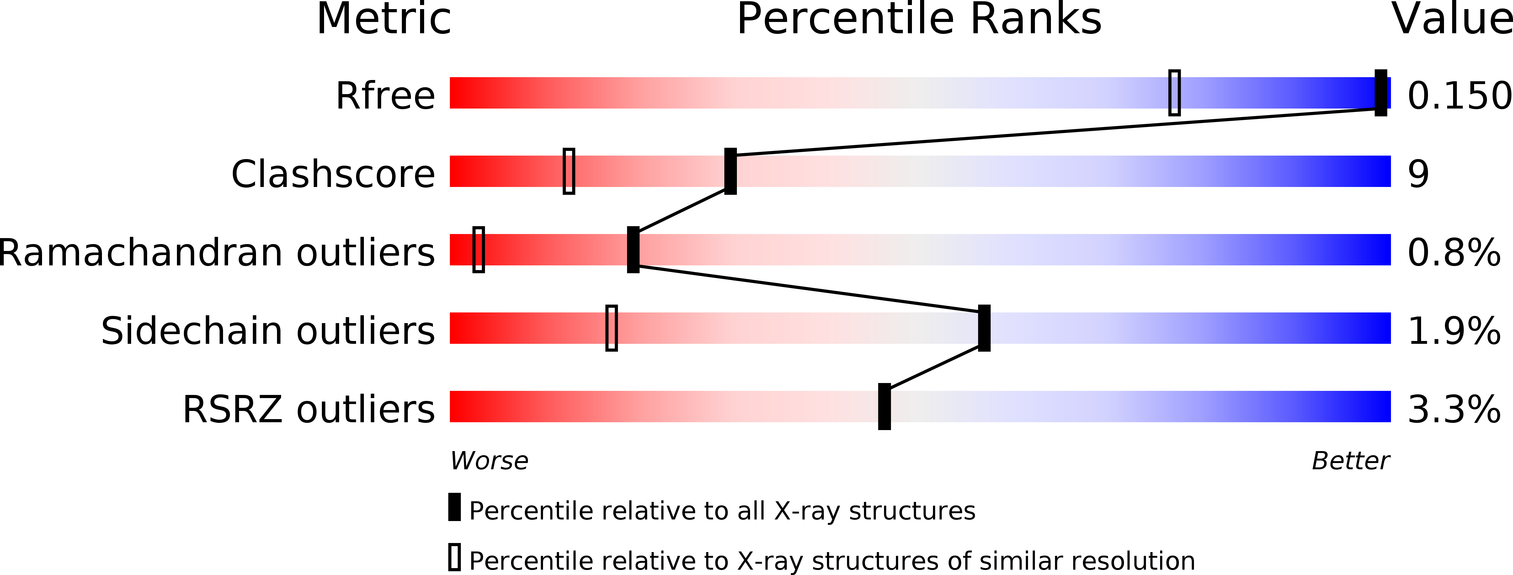
R-Value Free:
0.15
R-Value Work:
0.10
R-Value Observed:
0.10
Space Group:
P 21 21 21