3U7U image
Deposition Date 2011-10-14
Release Date 2012-08-29
Last Version Date 2024-11-27
Entry Detail
PDB ID:
3U7U
Title:
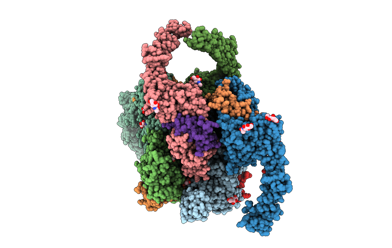
Crystal structure of extracellular region of human epidermal growth factor receptor 4 in complex with neuregulin-1 beta
Biological Source:
Source Organism:
Homo sapiens (Taxon ID: 9606)
Host Organism:
Method Details:
Experimental Method:
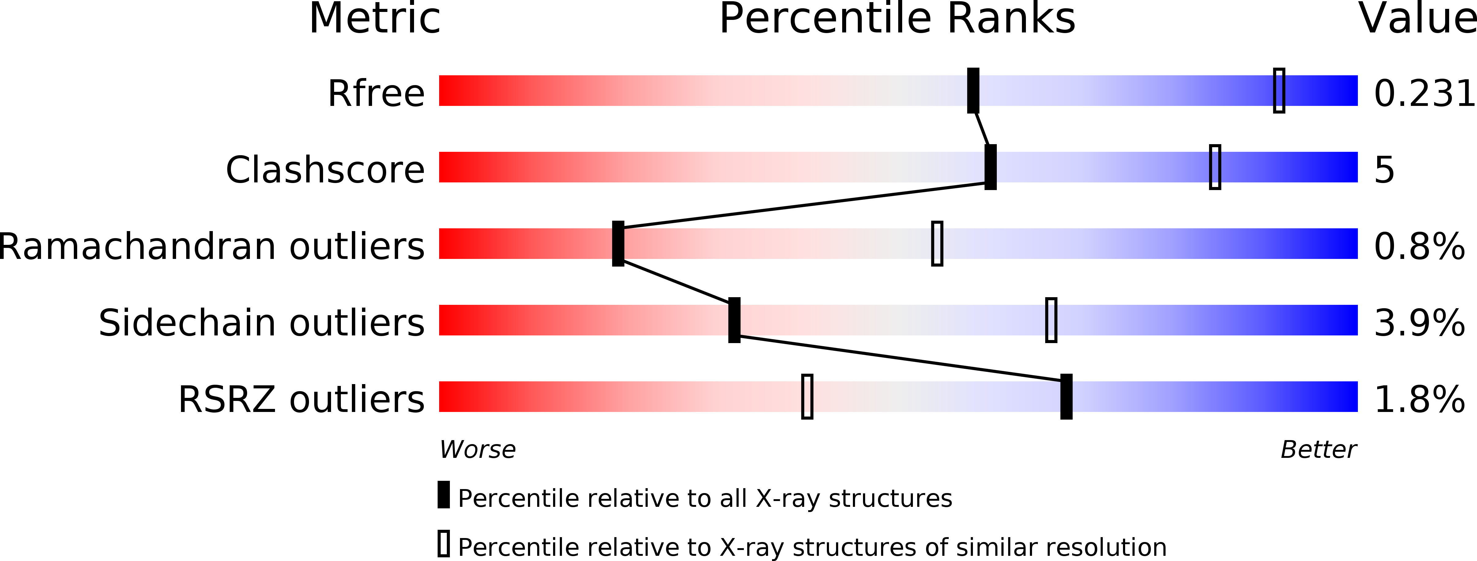
Resolution:
3.03 Å
R-Value Free:
0.22
R-Value Work:
0.18
R-Value Observed:
0.19
Space Group:
P 1 21 1
Macromolecular Entities
Polymer Type:polypeptide(L)
Molecule:Receptor tyrosine-protein kinase erbB-4
Gene (Uniprot):ERBB4
Chain IDs:A, B, C, D, E, F
Chain Length:615
Number of Molecules:6
Biological Source:Homo sapiens
Polymer Type:polypeptide(L)
Molecule:Neuregulin 1
Chain IDs:G, H, I, J, K, L
Chain Length:55
Number of Molecules:6
Biological Source:Homo sapiens
Modified Residue
Compound ID Chain ID Parent Comp ID Details 2D Image
ASN D ASN GLYCOSYLATION SITE
Ligand Molecules
Primary Citation
A single ligand is sufficient to activate EGFR dimers.
Proc.Natl.Acad.Sci.USA 109 10861 10866 (2012)
PMID: 22699492 DOI: 10.1073/pnas.1201114109

Abstact

Crystal structures of human epidermal growth factor receptor (EGFR) with bound ligand revealed symmetric, doubly ligated receptor dimers thought to represent physiologically active states. Such complexes fail to rationalize negative cooperativity of epidermal growth factor (EGF) binding to EGFR and the behavior of the ligandless EGFR homolog ErbB2/HER2, however. We report cell-based assays that provide evidence for active, singly ligated dimers of human EGFR and its homolog, ErbB4/HER4. We also report crystal structures of the ErbB4/HER4 extracellular region complexed with its ligand Neuregulin-1β that resolve two types of ErbB dimer when compared to EGFR:Ligand complexes. One type resembles the recently reported asymmetric dimer of Drosophila EGFR with a single high-affinity ligand bound and provides a model for singly ligated human ErbB dimers. These results unify models of vertebrate and invertebrate EGFR/ErbB signaling, imply that the tethered conformation of unliganded ErbBs evolved to prevent crosstalk among ErbBs, and establish a molecular basis for both negative cooperativity of ligand binding to vertebrate ErbBs and the absence of active ErbB2/HER2 homodimers in normal conditions.

Legend

Protein

Chemical

Disease

Primary Citation of related structures