3U6L image
Deposition Date 2011-10-12
Release Date 2012-04-25
Last Version Date 2024-10-30
Entry Detail
PDB ID:
3U6L
Keywords:
Title:
MutM set 2 CpGo
Biological Source:
Source Organism:
Host Organism:
Method Details:
Experimental Method:
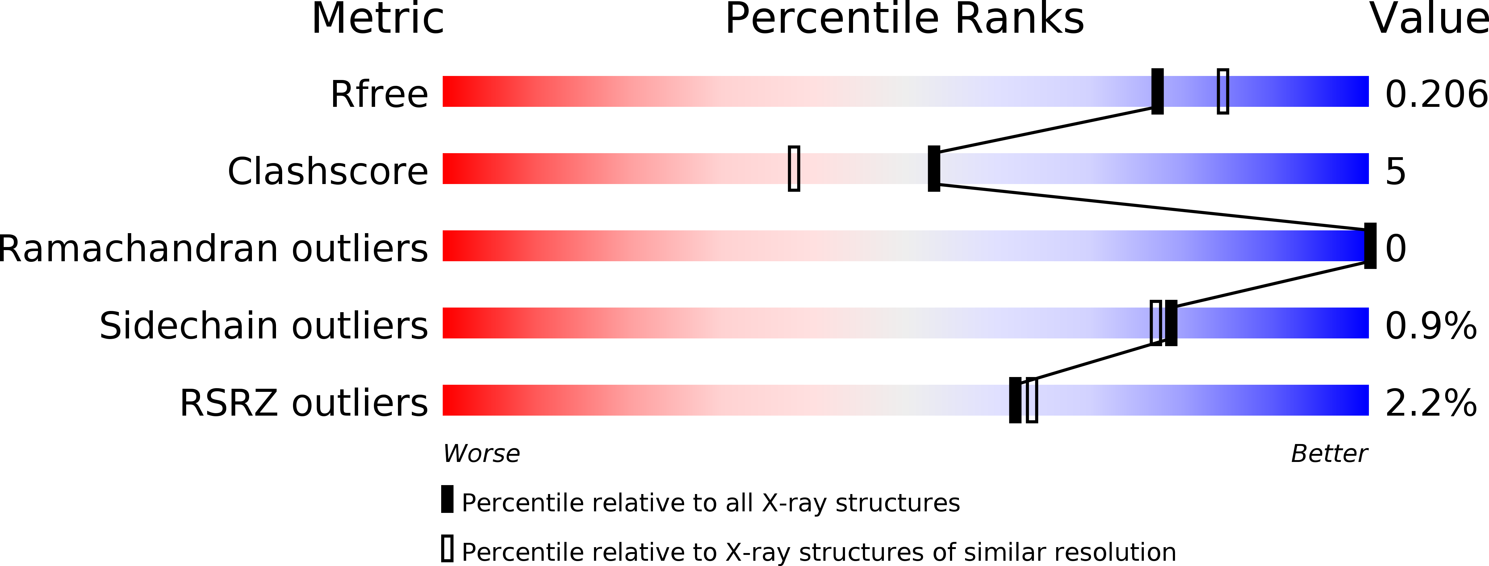
Resolution:
1.97 Å
R-Value Free:
0.20
R-Value Work:
0.17
R-Value Observed:
0.18
Space Group:
P 21 21 21
Macromolecular Entities
Structures with similar UniProt ID
Protein Blast
Polymer Type:polypeptide(L)
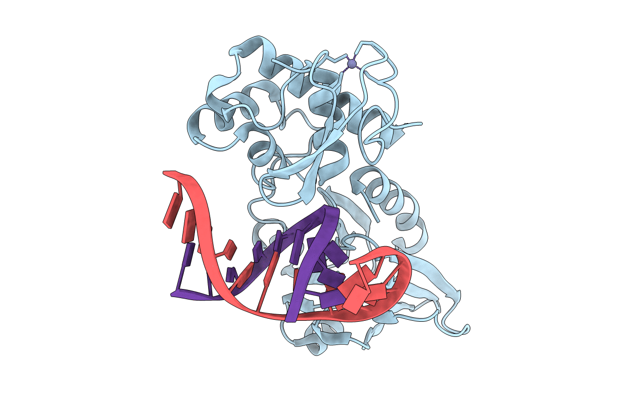
Molecule:Formamidopyrimidine-DNA glycosylase
Gene (Uniprot):mutM
Mutagens:E3Q, Q166C, V222P
Chain IDs:A
Chain Length:273
Number of Molecules:1
Biological Source:Geobacillus stearothermophilus
Polymer Type:polydeoxyribonucleotide
Molecule:DNA (5'-D(*AP*GP*GP*TP*AP*GP*AP*CP*CP*GP*GP*AP*CP*GP*C)-3')
Chain IDs:B
Chain Length:16
Number of Molecules:1
Biological Source:
Polymer Type:polydeoxyribonucleotide
Molecule:DNA (5'-D(*TP*GP*CP*GP*TP*CP*CP*(8OG)P*GP*TP*(CX2)P*TP*AP*CP*C)-3')
Chain IDs:C
Chain Length:16
Number of Molecules:1
Biological Source:
Modified Residue
Compound ID Chain ID Parent Comp ID Details 2D Image
8OG C DG ?
CX2 C DC ?
Ligand Molecules
Primary Citation
Sequence-dependent structural variation in DNA undergoing intrahelical inspection by the DNA glycosylase MutM.
J.Biol.Chem. 287 18044 18054 (2012)
PMID: 22465958 DOI: 10.1074/jbc.M111.313635

Abstact

MutM, a bacterial DNA-glycosylase, plays a critical role in maintaining genome integrity by catalyzing glycosidic bond cleavage of 8-oxoguanine (oxoG) lesions to initiate base excision DNA repair. The task faced by MutM of locating rare oxoG residues embedded in an overwhelming excess of undamaged bases is especially challenging given the close structural similarity between oxoG and its normal progenitor, guanine (G). MutM actively interrogates the DNA to detect the presence of an intrahelical, fully base-paired oxoG, whereupon the enzyme promotes extrusion of the target nucleobase from the DNA duplex and insertion into the extrahelical active site. Recent structural studies have begun to provide the first glimpse into the protein-DNA interactions that enable MutM to distinguish an intrahelical oxoG from G; however, these initial studies left open the important question of how MutM can recognize oxoG residues embedded in 16 different neighboring sequence contexts (considering only the 5'- and 3'-neighboring base pairs). In this study we set out to understand the manner and extent to which intrahelical lesion recognition varies as a function of the 5'-neighbor. Here we report a comprehensive, systematic structural analysis of the effect of the 5'-neighboring base pair on recognition of an intrahelical oxoG lesion. These structures reveal that MutM imposes the same extrusion-prone ("extrudogenic") backbone conformation on the oxoG lesion irrespective of its 5'-neighbor while leaving the rest of the DNA relatively free to adjust to the particular demands of individual sequences.

Legend

Protein

Chemical

Disease

Primary Citation of related structures