3U66 image
Deposition Date 2011-10-12
Release Date 2012-03-07
Last Version Date 2024-11-27
Entry Detail
PDB ID:
3U66
Title:
Crystal structure of T6SS SciP/TssL from Escherichia Coli Enteroaggregative 042
Biological Source:
Source Organism:
Host Organism:
Method Details:
Experimental Method:
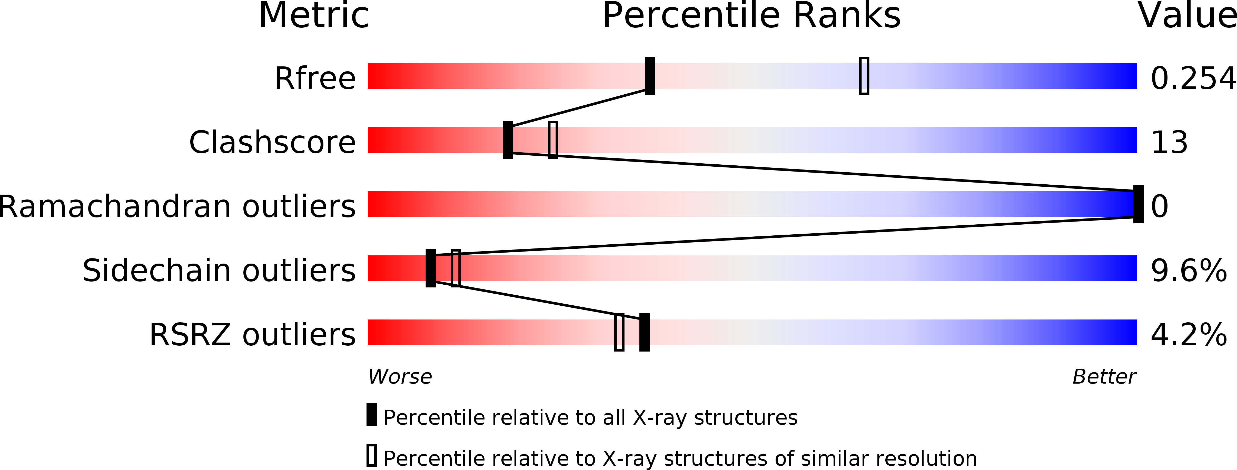
Resolution:
2.63 Å
R-Value Free:
0.25
R-Value Work:
0.23
R-Value Observed:
0.23
Space Group:
I 2 2 2
Macromolecular Entities
Polymer Type:polypeptide(L)
Molecule:Putative type VI secretion protein
Gene (Uniprot):EC042_4527
Chain IDs:A
Chain Length:183
Number of Molecules:1
Biological Source:Escherichia coli
Modified Residue
Compound ID Chain ID Parent Comp ID Details 2D Image
MSE A MET SELENOMETHIONINE
Ligand Molecules
Primary Citation
Structural characterization and oligomerization of the TssL protein, a component shared by bacterial type VI and type IVb secretion systems.
J.Biol.Chem. 287 14157 14168 (2012)
PMID: 22371492 DOI: 10.1074/jbc.M111.338731

Abstact

The Type VI secretion system (T6SS) is a macromolecular system distributed in Gram-negative bacteria, responsible for the secretion of effector proteins into target cells. The T6SS has a broad versatility as it can target both eukaryotic and prokaryotic cells. It is therefore involved in host pathogenesis or killing neighboring bacterial cells to colonize a new niche. At the architecture level, the T6SS core apparatus is composed of 13 proteins, which assemble in two subcomplexes. One of these subcomplexes, composed of subunits that share structural similarities with bacteriophage tail and baseplate components, is anchored to the cell envelope by the membrane subcomplex. This latter is constituted of at least three proteins, TssL, TssM, and TssJ. The crystal structure of the TssJ outer membrane lipoprotein and its interaction with the inner membrane TssM protein have been recently reported. TssL and TssM share sequence homology and characteristics with two components of the Type IVb secretion system (T4bSS), IcmH/DotU and IcmF, respectively. In this study, we report the crystal structure of the cytoplasmic domain of the TssL inner membrane protein from the enteroaggregative Escherichia coli Sci-1 T6SS. It folds as a hook-like structure composed of two three-helix bundles. Two TssL molecules associate to form a functional complex. Although the TssL trans-membrane segment is the main determinant of self-interaction, contacts between the cytoplasmic domains are required for TssL function. Based on sequence homology and secondary structure prediction, we propose that the TssL structure is the prototype for the members of the TssL and IcmH/DotU families.

Legend

Protein

Chemical

Disease

Primary Citation of related structures