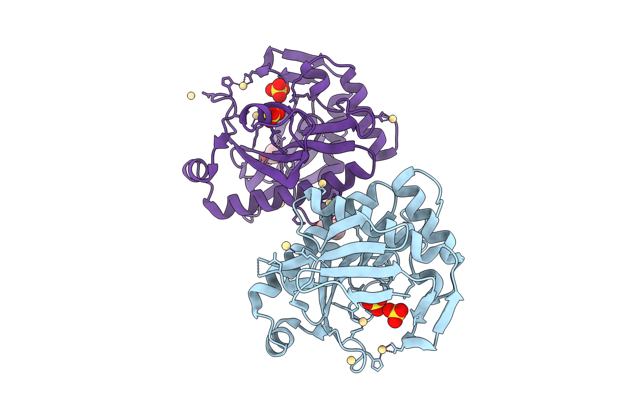
3U54 image
Deposition Date 2011-10-11
Release Date 2012-10-17
Last Version Date 2024-11-13
Entry Detail
PDB ID:
3U54
Keywords:
Title:
Crystal structure (Type-1) of SAICAR synthetase from Pyrococcus horikoshii OT3
Biological Source:
Source Organism:
Host Organism:
Method Details:
Experimental Method:
Resolution:
2.35 Å
R-Value Free:
0.28
R-Value Work:
0.23
R-Value Observed:
0.23
Space Group:
H 3
Macromolecular Entities
Structures with similar UniProt ID
Protein Blast
Polymer Type:polypeptide(L)
Molecule:Phosphoribosylaminoimidazole-succinocarboxamide synthase
Gene (Uniprot):purC
Chain IDs:A, B
Chain Length:238
Number of Molecules:2
Biological Source:Pyrococcus horikoshii
Modified Residue
Compound ID Chain ID Parent Comp ID Details 2D Image
MSE A MET SELENOMETHIONINE
Primary Citation
Structure of SAICAR synthetase from Pyrococcus horikoshii OT3: insights into thermal stability
Int.J.Biol.Macromol. 53 7 19 (2013)
PMID: 23137517 DOI: 10.1016/j.ijbiomac.2012.10.028

Abstact

The first native crystal structure of Phosphoribosylaminoimidazole-succinocarboxamide synthetase (SAICAR synthetase) from a hyperthermophilic organism Pyrococcus horikoshii OT3 was determined in two space groups H3 (Type-1: Resolution 2.35Å) and in C222(1) (Type-2: Resolution 1.9Å). Both are dimeric but Type-1 structure exhibited hexameric arrangement due to the presence of cadmium ions. A comparison has been made on the sequence and structures of all SAICAR synthetases to better understand the differences between mesophilic, thermophilic and hyperthermophilic SAICAR synthetases. These SAICAR synthetases are reasonably similar in sequence and three-dimensional structure; however, differences were visible only in the subtler details of percentage composition of the sequences, salt bridge interactions and non-polar contact areas.

Legend

Protein

Chemical

Disease

Primary Citation of related structures