Deposition Date
2011-10-09
Release Date
2012-04-25
Last Version Date
2024-10-16
Entry Detail
PDB ID:
3U4L
Keywords:
Title:
Cryocooled bovine profilin:actin crystal structure to 2.4 A
Biological Source:
Source Organism(s):
Bos taurus (Taxon ID: 9913)
Method Details:
Experimental Method:
Resolution:
2.40 Å
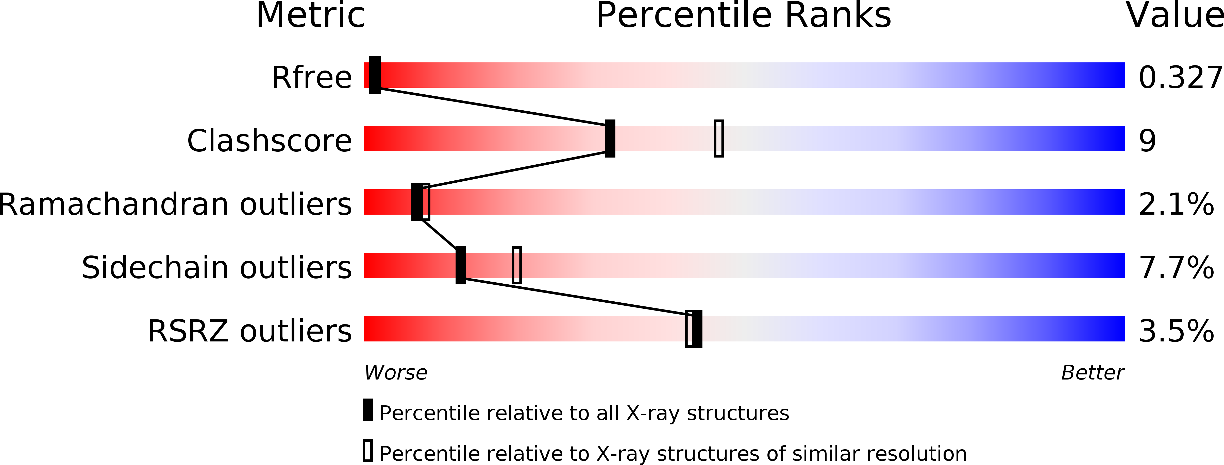
R-Value Free:
0.33
R-Value Work:
0.24
R-Value Observed:
0.24
Space Group:
P 21 21 21