Deposition Date
2011-10-09
Release Date
2012-09-26
Last Version Date
2024-11-20
Entry Detail
PDB ID:
3U4K
Keywords:
Title:
Crystal structure of the receptor binding domain of plasmid-born adhesin MrkD1P of Klebsiella pneumoniae
Biological Source:
Source Organism(s):
Klebsiella pneumoniae (Taxon ID: 573)
Expression System(s):
Method Details:
Experimental Method:
Resolution:
3.00 Å
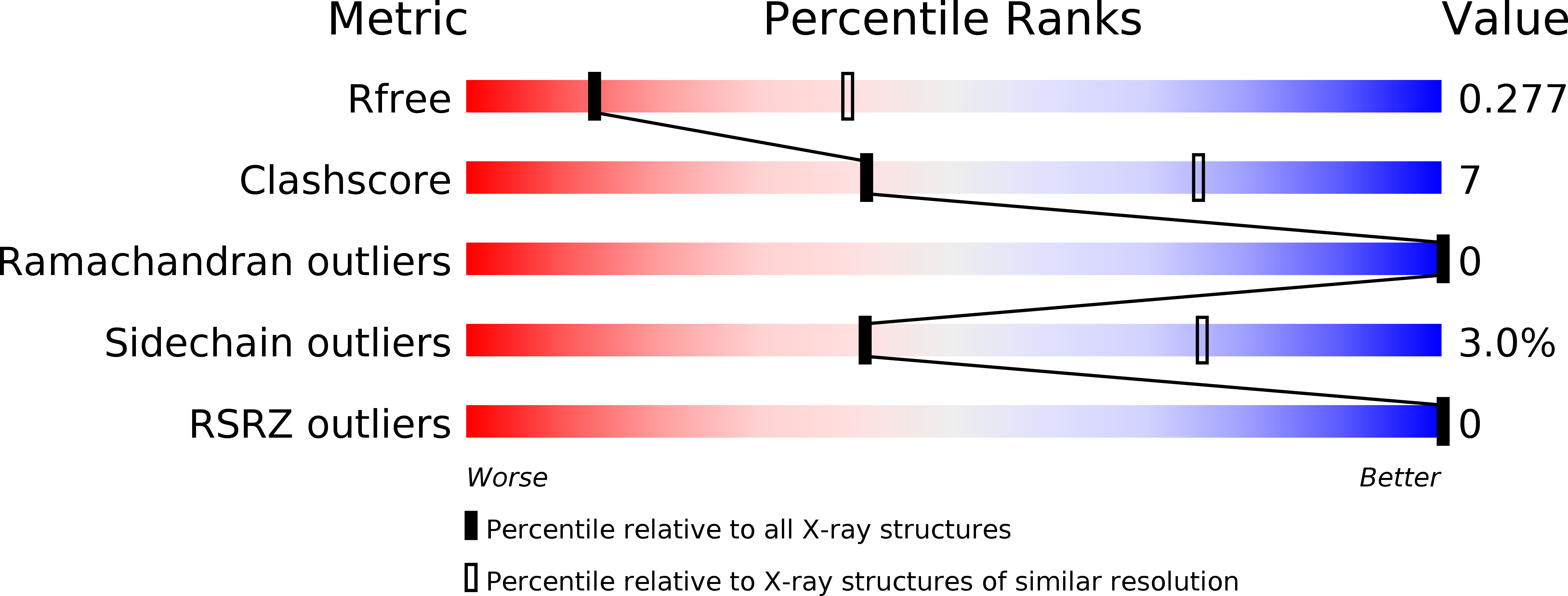
R-Value Free:
0.27
R-Value Work:
0.22
R-Value Observed:
0.22
Space Group:
P 31 2 1