3U3W image
Deposition Date 2011-10-06
Release Date 2013-01-09
Last Version Date 2023-09-13
Entry Detail
PDB ID:
3U3W
Title:
Crystal Structure of Bacillus thuringiensis PlcR in complex with the peptide PapR7 and DNA
Biological Source:
Source Organism:
Host Organism:
Method Details:
Experimental Method:
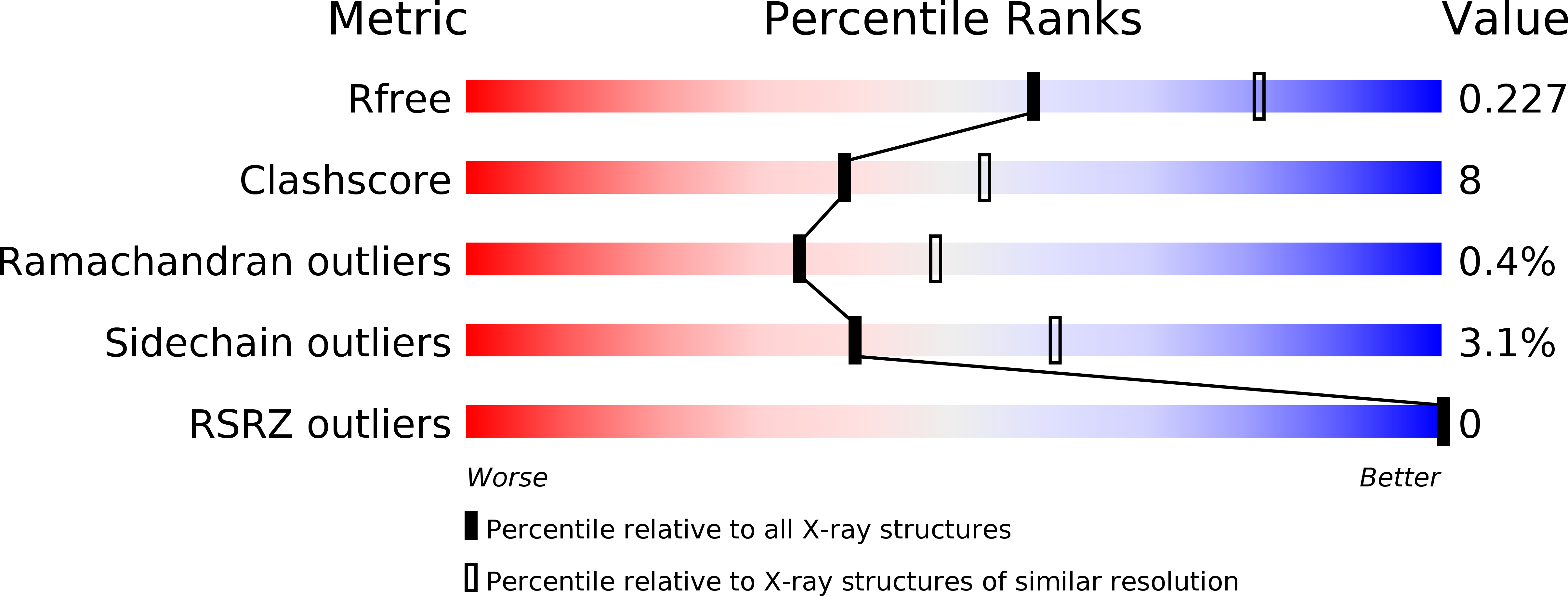
Resolution:
2.40 Å
R-Value Free:
0.23
R-Value Work:
0.17
R-Value Observed:
0.18
Space Group:
P 1 21 1
Macromolecular Entities
Polymer Type:polypeptide(L)
Molecule:Transcriptional activator PlcR protein
Gene (Uniprot):plcR
Chain IDs:A, B
Chain Length:293
Number of Molecules:2
Biological Source:Bacillus thuringiensis
Polymer Type:polypeptide(L)
Molecule:C-terminus heptapeptide from PapR protein
Gene (Uniprot):papR
Chain IDs:C (auth: P), D (auth: Q)
Chain Length:7
Number of Molecules:2
Biological Source:Bacillus cereus
Ligand Molecules
Primary Citation
Structural basis for the activation mechanism of the PlcR virulence regulator by the quorum-sensing signal peptide PapR.
Proc.Natl.Acad.Sci.USA 110 1047 1052 (2013)
PMID: 23277548 DOI: 10.1073/pnas.1213770110

Abstact

The quorum-sensing regulator PlcR is the master regulator of most known virulence factors in Bacillus cereus. It is a helix-turn-helix (HTH)-type transcription factor activated upon binding of its cognate signaling peptide PapR on a tetratricopeptide repeat-type regulatory domain. The structural and functional properties of PlcR have defined a new family of sensor regulators, called the RNPP family (for Rap, NprR, PrgX, and PlcR), in Gram-positive bacteria. To fully understand the activation mechanism of PlcR, we took a closer look at the conformation changes induced upon binding of PapR and of its target DNA, known as PlcR-box. For that purpose we have determined the structures of the apoform of PlcR (Apo PlcR) and of the ternary complex of PlcR with PapR and the PlcR-box from the plcA promoter. Comparison of the apoform of PlcR with the previously published structure of the PlcR-PapR binary complex shows how a small conformational change induced in the C-terminal region of the tetratricopeptide repeat (TPR) domain upon peptide binding propagates via the linker helix to the N-terminal HTH DNA-binding domain. Further comparison with the PlcR-PapR-DNA ternary complex shows how the activation of the PlcR dimer allows the linker helix to undergo a drastic conformational change and subsequent proper positioning of the HTH domains in the major groove of the two half sites of the pseudopalindromic PlcR-box. Together with random mutagenesis experiments and interaction measurements using peptides from distinct pherogroups, this structural analysis allows us to propose a molecular mechanism for this functional switch.

Legend

Protein

Chemical

Disease

Primary Citation of related structures