3U3V image
Deposition Date 2011-10-06
Release Date 2012-05-02
Last Version Date 2024-11-06
Entry Detail
PDB ID:
3U3V
Keywords:
Title:
The S-SAD phased crystal structure of the ecto-domain of Death Receptor 6 (DR6)
Biological Source:
Source Organism:
Homo sapiens (Taxon ID: 9606)
Host Organism:
Method Details:
Experimental Method:
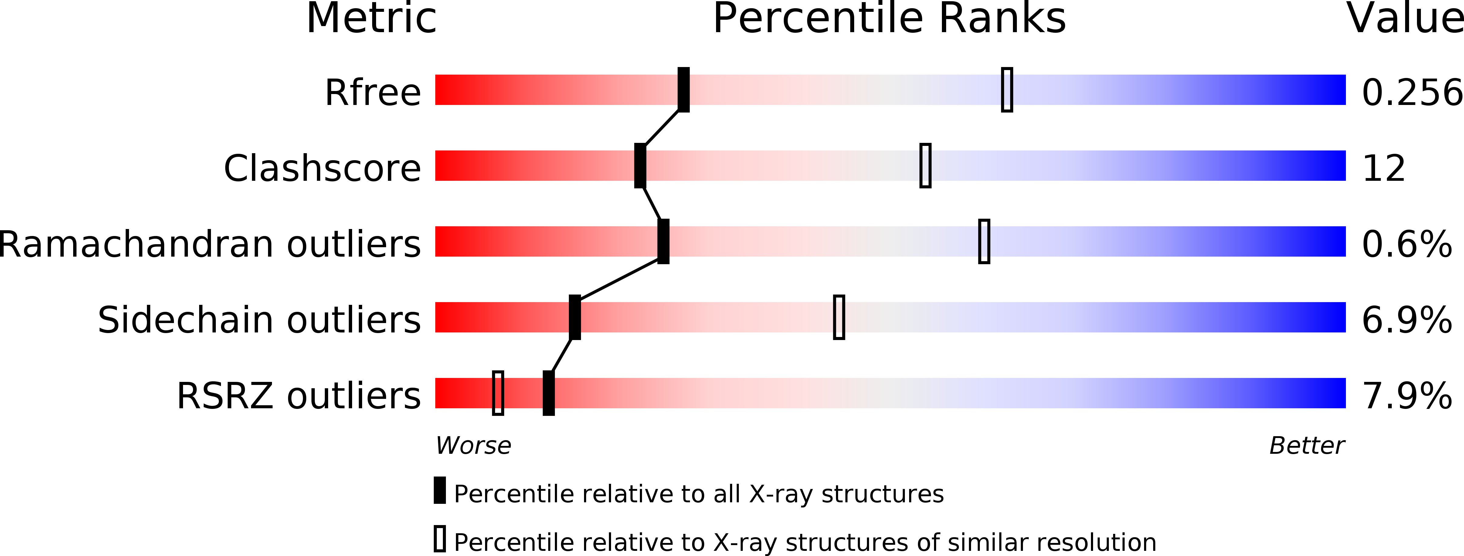
Resolution:
2.96 Å
R-Value Free:
0.25
R-Value Work:
0.19
R-Value Observed:
0.19
Space Group:
P 61 2 2
Macromolecular Entities
Polymer Type:polypeptide(L)
Molecule:Tumor necrosis factor receptor superfamily member 21
Gene (Uniprot):TNFRSF21
Chain IDs:A
Chain Length:313
Number of Molecules:1
Biological Source:Homo sapiens
Primary Citation
S-SAD phasing study of death receptor 6 and its solution conformation revealed by SAXS
Acta Crystallogr.,Sect.D 68 521 530 (2012)
PMID: 22525750 DOI: 10.1107/S0907444912004490

Abstact

A subset of tumour necrosis factor receptor (TNFR) superfamily members contain death domains in their cytoplasmic tails. Death receptor 6 (DR6) is one such member and can trigger apoptosis upon the binding of a ligand by its cysteine-rich domains (CRDs). The crystal structure of the ectodomain (amino acids 1-348) of human death receptor 6 (DR6) encompassing the CRD region was phased using the anomalous signal from S atoms. In order to explore the feasibility of S-SAD phasing at longer wavelengths (beyond 2.5 Å), a comparative study was performed on data collected at wavelengths of 2.0 and 2.7 Å. In spite of sub-optimal experimental conditions, the 2.7 Å wavelength used for data collection showed potential for S-SAD phasing. The results showed that the R(ano)/R(p.i.m.) ratio is a good indicator for monitoring the anomalous data quality when the anomalous signal is relatively strong, while d''/sig(d'') calculated by SHELXC is a more sensitive and stable indicator applicable for grading a wider range of anomalous data qualities. The use of the `parameter-space screening method' for S-SAD phasing resulted in solutions for data sets that failed during manual attempts. SAXS measurements on the ectodomain suggested that a dimer defines the minimal physical unit of an unliganded DR6 molecule in solution.

Legend

Protein

Chemical

Disease

Primary Citation of related structures