3U2P image
Deposition Date 2011-10-04
Release Date 2011-11-09
Last Version Date 2024-11-20
Entry Detail
PDB ID:
3U2P
Title:
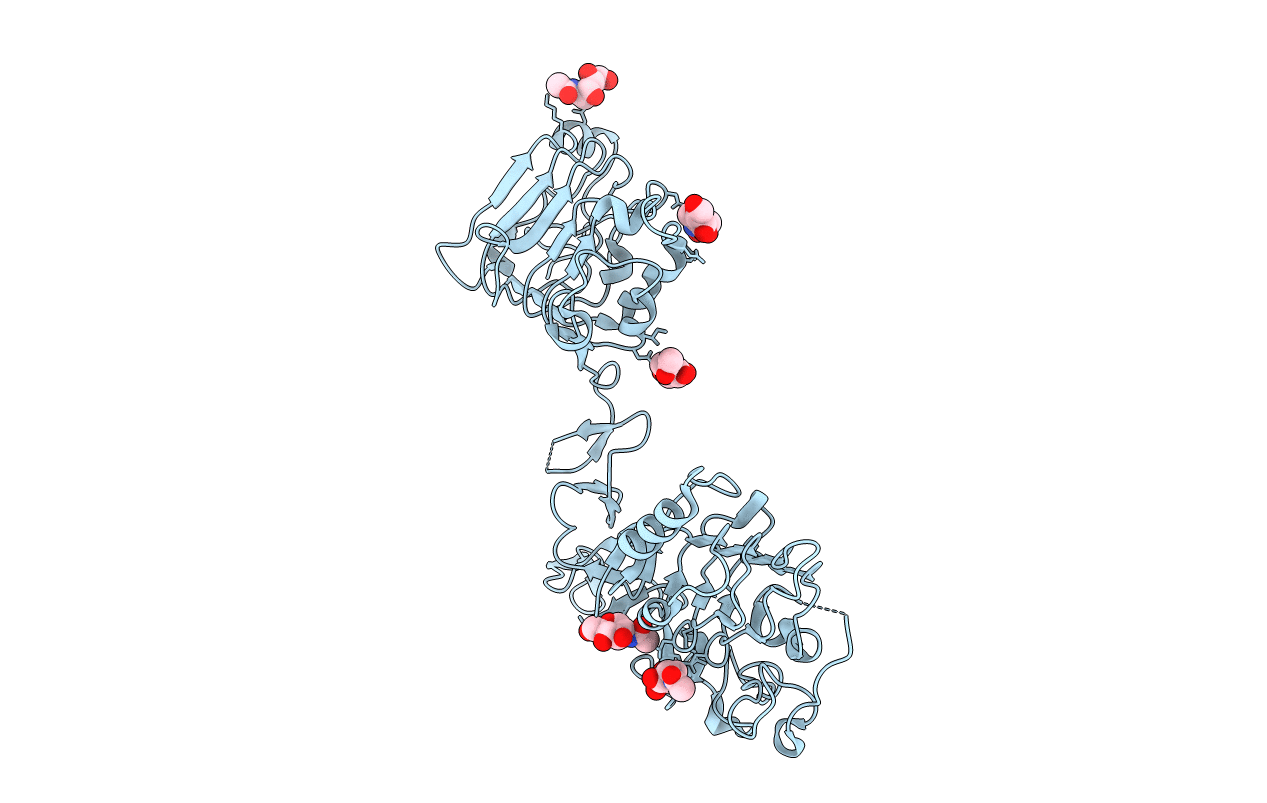
Crystal structure of N-terminal three extracellular domains of ErbB4/Her4
Biological Source:
Source Organism:
Homo sapiens (Taxon ID: 9606)
Host Organism:
Method Details:
Experimental Method:
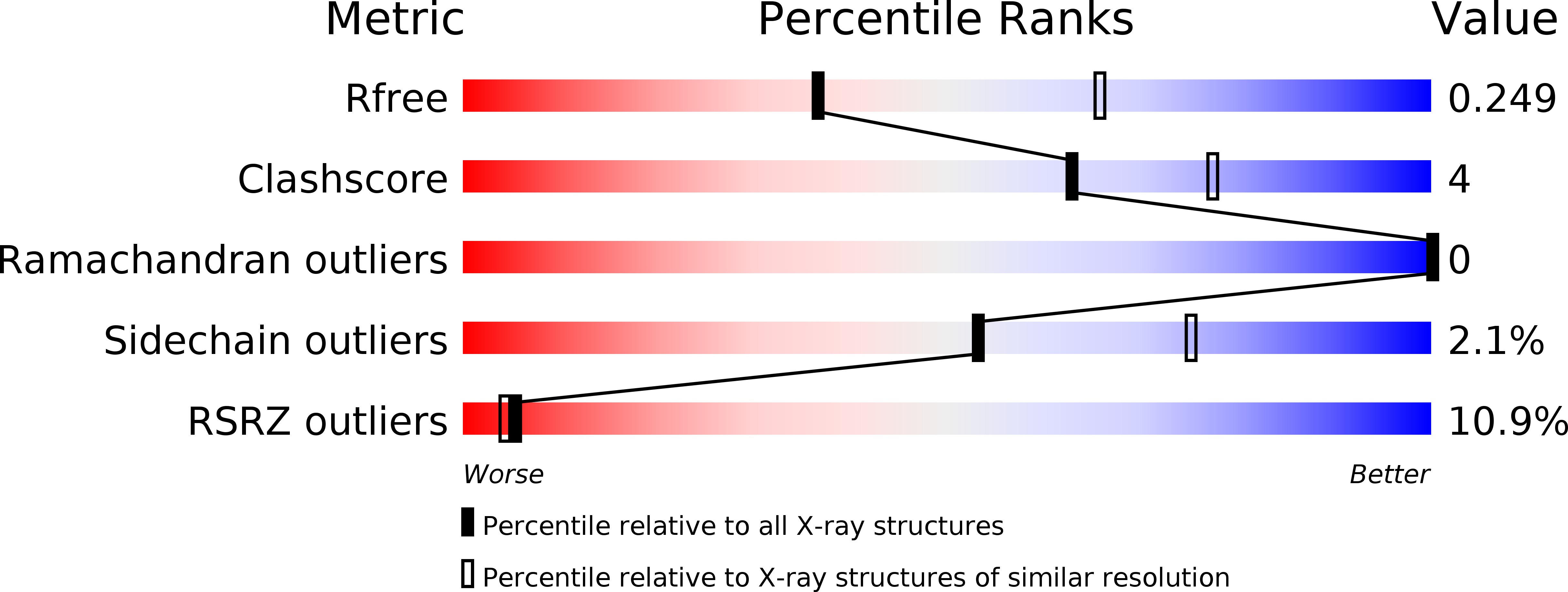
Resolution:
2.57 Å
R-Value Free:
0.24
R-Value Work:
0.19
R-Value Observed:
0.19
Space Group:
P 21 21 21
Macromolecular Entities
Polymer Type:polypeptide(L)
Molecule:Receptor tyrosine-protein kinase erbB-4
Gene (Uniprot):ERBB4
Chain IDs:A
Chain Length:497
Number of Molecules:1
Biological Source:Homo sapiens
Modified Residue
Compound ID Chain ID Parent Comp ID Details 2D Image
ASN A ASN GLYCOSYLATION SITE
Ligand Molecules
Primary Citation
The ErbB4 extracellular region retains a tethered-like conformation in the absence of the tether.
Protein Sci. 21 152 155 (2012)
PMID: 22012915 DOI: 10.1002/pro.753

Abstact

The epidermal growth factor receptor (EGFR) and its homologs ErbB3 and ErbB4 adopt a tethered conformation in the absence of ligand in which an extended hairpin loop from domain II contacts the juxtamembrane region of domain IV and tethers the domain I/II pair to the domain III/IV pair. By burying the hairpin loop, which is required for formation of active receptor dimers, the tether contact was thought to prevent constitutive activation of EGFR and its homologs. Amino-acid substitutions at key sites within the tether contact region fail to result in constitutively active receptors however. We report here the 2.5 Å crystal structure of the N-terminal three extracellular domains of ErbB4, which bind ligand but lack domain IV and thus the tether contact. This ErbB4 fragment nonetheless adopts a domain arrangement very similar to the arrangement adopted in the presence of the tether suggesting that regions in addition to the tether contribute to maintaining this conformation and inactivity in the absence of the tether contact. We suggest that the tether conformation may have evolved to prevent crosstalk between different EGFR homologs and thus allow diversification of EGFR and its homologs.

Legend

Protein

Chemical

Disease

Primary Citation of related structures