3U2F image
Entry Detail
PDB ID:
3U2F
Title:
ATP synthase c10 ring in proton-unlocked conformation at PH 8.3
Biological Source:
Source Organism:
PDB Version:
Deposition Date:
2011-10-03
Release Date:
2012-02-08
Method Details:
Experimental Method:
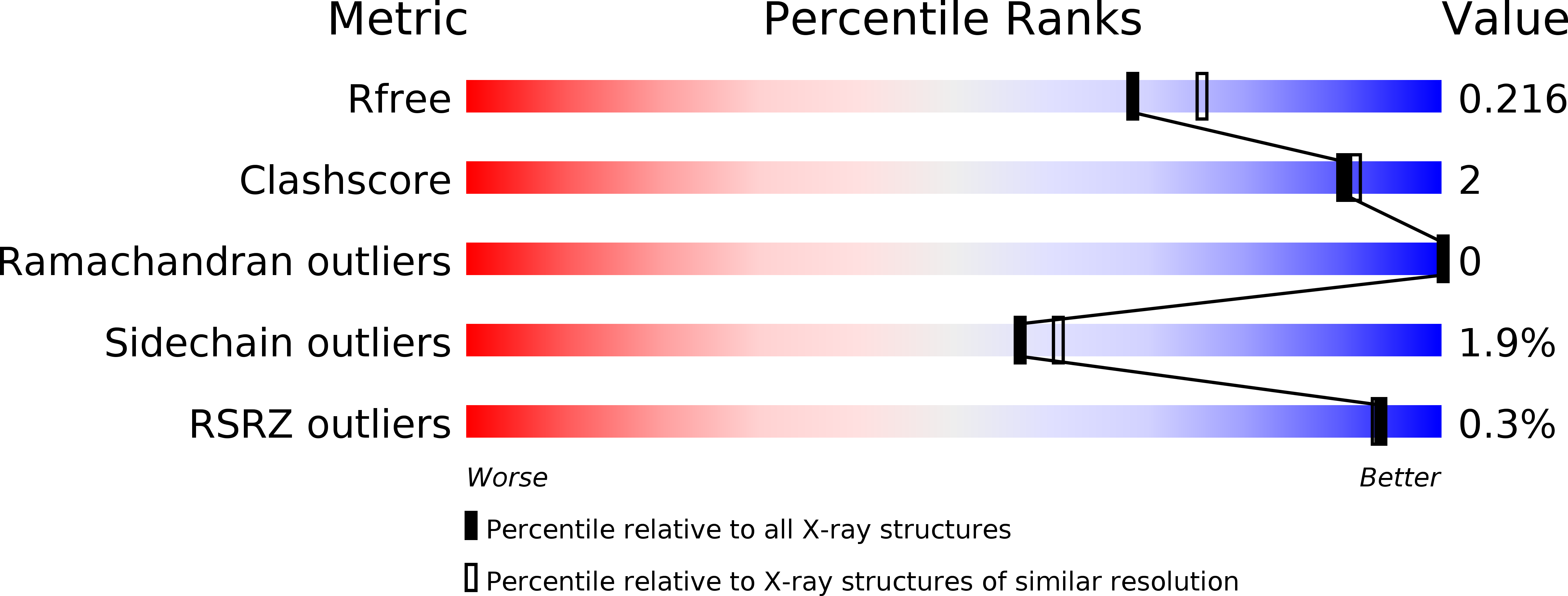
Resolution:
2.00 Å
R-Value Free:
0.21
R-Value Work:
0.19
R-Value Observed:
0.19
Space Group:
P 42 2 2
Macromolecular Entities
Polymer Type:polypeptide(L)
Description:ATP synthase subunit C, mitochondrial
Chain IDs:A (auth: K), B (auth: L), C (auth: M), D (auth: N), E (auth: O)
Chain Length:76
Number of Molecules:5
Biological Source:Saccharomyces cerevisiae
Modified Residue
Compound ID Chain ID Parent Comp ID Details 2D Image
FME A MET N-FORMYLMETHIONINE
Primary Citation
Structure of the c(10) ring of the yeast mitochondrial ATP synthase in the open conformation.
Nat.Struct.Mol.Biol. 19 485 491 (2012)
PMID: 22504883 DOI: 10.1038/nsmb.2284

Abstact

The proton pore of the F(1)F(o) ATP synthase consists of a ring of c subunits, which rotates, driven by downhill proton diffusion across the membrane. An essential carboxylate side chain in each subunit provides a proton-binding site. In all the structures of c-rings reported to date, these sites are in a closed, ion-locked state. Structures are here presented of the c(10) ring from Saccharomyces cerevisiae determined at pH 8.3, 6.1 and 5.5, at resolutions of 2.0 Å, 2.5 Å and 2.0 Å, respectively. The overall structure of this mitochondrial c-ring is similar to known homologs, except that the essential carboxylate, Glu59, adopts an open extended conformation. Molecular dynamics simulations reveal that opening of the essential carboxylate is a consequence of the amphiphilic nature of the crystallization buffer. We propose that this new structure represents the functionally open form of the c subunit, which facilitates proton loading and release.

Legend

Protein

Chemical

Disease

Primary Citation of related structures