
Deposition Date
2011-09-30
Release Date
2011-11-23
Last Version Date
2024-10-16
Entry Detail
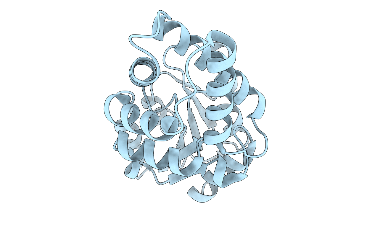
PDB ID:
3U26
Keywords:
Title:
Crystal Structure of Engineered Protein. Northeast Structural Genomics Consortium Target OR48
Biological Source:
Source Organism(s):
Pyrococcus horikoshii (Taxon ID: 53953)
Expression System(s):
Method Details:
Experimental Method:
Resolution:
1.59 Å
R-Value Free:
0.24
R-Value Work:
0.19
R-Value Observed:
0.20
Space Group:
P 1 21 1
