3U1I image
Deposition Date 2011-09-29
Release Date 2011-11-09
Last Version Date 2023-11-15
Entry Detail
PDB ID:
3U1I
Title:
Dengue virus protease covalently bound to a peptide
Biological Source:
Source Organism:
Dengue virus 3 (Taxon ID: 408693)
(Taxon ID: )
Host Organism:
Method Details:
Experimental Method:
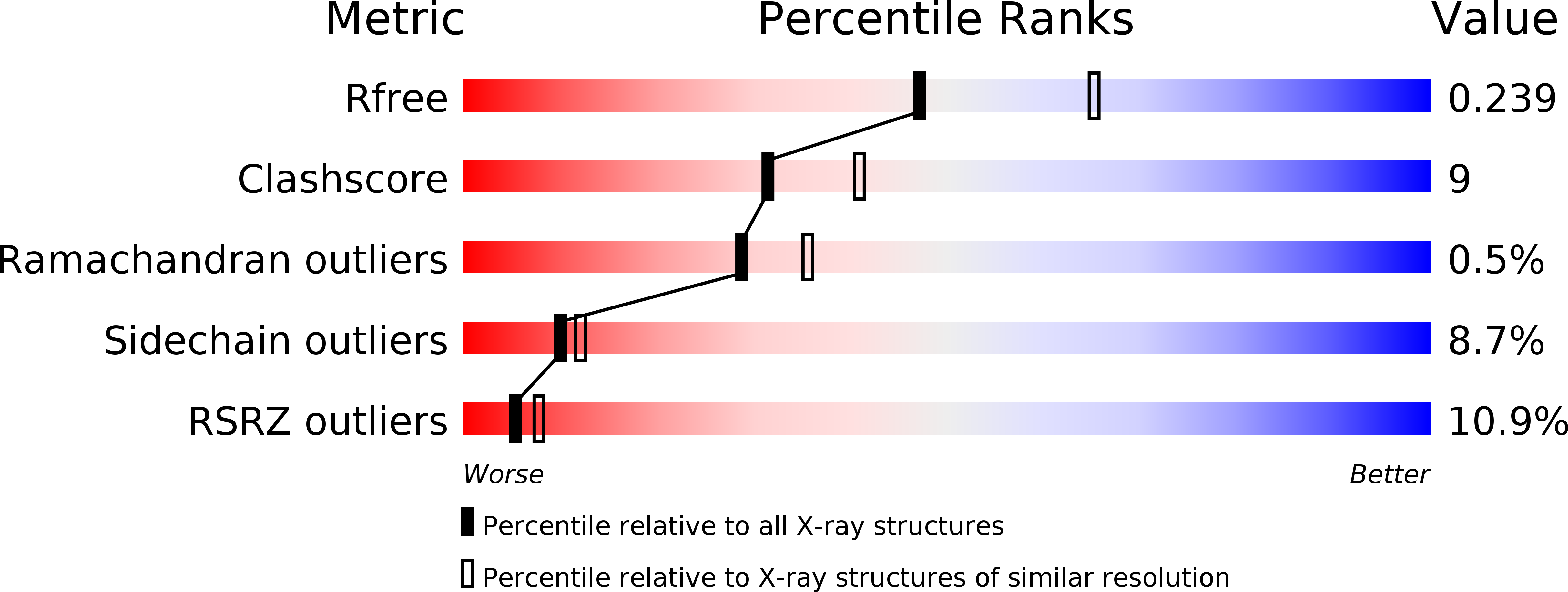
Resolution:
2.30 Å
R-Value Free:
0.24
R-Value Work:
0.21
R-Value Observed:
0.21
Space Group:
C 2 2 21
Macromolecular Entities
Polymer Type:polypeptide(L)
Molecule:Serine protease subunit NS2B
Chain IDs:A, C
Chain Length:51
Number of Molecules:2
Biological Source:Dengue virus 3
Polymer Type:polypeptide(L)
Molecule:Serine protease NS3
Chain IDs:B, D
Chain Length:191
Number of Molecules:2
Biological Source:Dengue virus 3
Polymer Type:polypeptide(L)
Molecule:peptide of (BEZ)(NLE)KR(OAR)
Chain IDs:E, F
Chain Length:5
Number of Molecules:2
Biological Source:
Modified Residue
Compound ID Chain ID Parent Comp ID Details 2D Image
NLE F LEU NORLEUCINE
OAR E ARG N-(4-AMINO-5-HYDROXY-PENTYL)-GUANIDINE
Ligand Molecules
Peptide-like Molecules
PRD_000988
Primary Citation
Ligand-bound structures of the dengue virus protease reveal the active conformation
J.Virol. 86 438 446 (2012)
PMID: 22031935 DOI: 10.1128/JVI.06225-11

Abstact

Dengue is a mosquito-borne viral hemorrhagic disease that is a major threat to human health in tropical and subtropical regions. Here we report crystal structures of a peptide covalently bound to dengue virus serotype 3 (DENV-3) protease as well as the serine-protease inhibitor aprotinin bound to the same enzyme. These structures reveal, for the first time, a catalytically active, closed conformation of the DENV protease. In the presence of the peptide, the DENV-3 protease forms the closed conformation in which the hydrophilic β-hairpin region of NS2B wraps around the NS3 protease core, in a manner analogous to the structure of West Nile virus (WNV) protease. Our results confirm that flavivirus proteases form the closed conformation during proteolysis, as previously proposed for WNV. The current DENV-3 protease structures reveal the detailed interactions at the P4' to P3 sites of the substrate. The new structural information explains the sequence preference, particularly for long basic residues in the nonprime side, as well as the difference in substrate specificity between the WNV and DENV proteases at the prime side. Structural analysis of the DENV-3 protease-peptide complex revealed a pocket that is formed by residues from NS2B and NS3; this pocket also exists in the WNV NS2B/NS3 protease structure and could be targeted for potential antivirus development. The structural information presented in the current study is invaluable for the design of specific inhibitors of DENV protease.

Legend

Protein

Chemical

Disease

Primary Citation of related structures